Download
1 / 27
290 likes | 444 Views
Explore the types and characteristics of stratified epithelium, including nonkeratinized and keratinized squamous cells, cuboidal, and columnar layers. Learn about transitional epithelium and its protective functions in the urinary system.

E N D
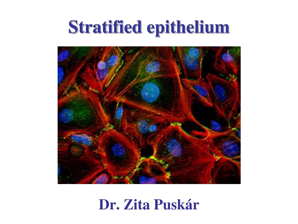
Stratified epithelium Dr. Zita Puskár
Stratified epithelium Nonkeratinized Stratified squamous Keratinized Stratified cuboidal Stratified columnar Transitional epithelium (urothelium) Classification is based on the cell shape of the superficial layer.
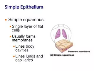
General features of stratified squamous epithelia keratinized nonkeratinized Flattened cells in the superficial layer Appear in areas where mechanical insults are frequent Fastness: stratification projections of the connective tissue (papillae) firm cytoskeleton (keratin filaments) firm intercellular junctions (connections) – desmosomes hemidesmosomes at the border of epithelium and connective tissue Avascular and nourished by diffusion of nutrients from capillaries in the connective tissue
Stratified squamousnonkeratinized epithelia nonkeratinized 1. Basal layer (stratum basale/germinativum) Basophil columnar cells with mitotoic activity, (epithelial stem cells are among them) → production of dividing and differentiating cells 2. Spinous layer (stratum spinosum/poligonale) Polyhedral cells with desmosomes (intermedier filaments: keratin filaments - bundles tonofilaments) During histological preparation the cells shrink but the desmosomal connections remain intact → spinous appearance 3. stratum planocellulare/superficiale The top layers consist of living flattened cells with elongated nuclei. 3 2 1
Oral cavity tongue
Cornea No connective tissue papillae!!!
Stratified squamous keratinized epithelium New differentiated cells (pushed upward) → keratinization (keratin-tonofibrill), lamellar granules → apoptosis (programmed cell death) → squames cornified cells (continuously shed) Layers: • Basal layer (Str. basale / germinativum) 2. Spinous layer (Stratum spinosum / poligonale) 3. Granular layer (Stratum granulosum) 3-5 cell layers, flattened poligonal cells Keratohyaline granules in the cytoplasm → basophil staining Lamellar granules (membrane coated lipids) → exocytosis → lipid envelope (barrier against water loss) • Stratum lucidum (seen only in thick skin)thin homogenous translucent eosinophilic layer. Nuclei and organelles are lost. • Stratum corneum Cells contain only fibrillar and amorphous proteins with thickened plasma membrane (4)-5 3 2 1 thin skin
Thick skin (palms and soles) • Str. basale / germinativum • Stratum spinosum / poligonale • Stratum granulosum • Stratum lucidum • Stratum corneum (continuously shed) Str. corneum 5 Str. lucidum 3 4 2 1
Keratinization Pachyonychia congenita: A rare genodermatosis.Agarwal P, Chhaperwal MK, Singh A, Verma A, Nijhawan M, Singh K, Mathur D - Indian Dermatol Online J (2013)
Keratinization https://www.youtube.com/watch?v=OKosGSm7Ps4 by Walter Jahn
A disease of desmosomes: pemphigus Blistering autoimmune disease in which antibodies form against desmoglein (the transmembrane desmosomal cadherin) and the cells of stratum spinosum are separated from each other (unglued). (Blisters→sores)
Cells of epidermis lipid layers lamellar body Keratinocyte (epithelial cell) Langerhans cells Merkel cells nerve ending Melanocyte Pigmented epithelial cell
Nobel-prize in Chemistry 2003 P. Agre and R. MacKinnon: water and ion channels
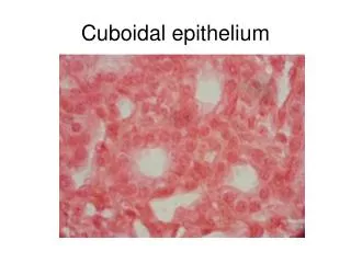
Stratified cuboidal epithelium Gland ducts Sweat gland
Stratified columnar epithelium Basal and superficial cells are columnar, poligonal cells in between. Large ducts of exocrine glands, spongy part of male urethra, fornix of conjunctiva
Transitional epithelium - urothelium umbrella cells pear-shaped cells Characteristic epithelium of urinary system. Transitional: distended bladder compress the cells into squamous cells, empty bladder results in thickening of the epithelium. Protections from hypertonic and cytotoxic effect of urine. Layers 1. basal layer: cuboidal or columnar epithelial cells with mitotic activity 2. piriform layer: poligonal parabasal cells – pear shaped cells 3. dome shaped-umbrella cell layeroften binucleated cells. Appearance: renal pelvis, ureter, urinary bladder, upper part of urethra
Umbrella cell plaques vesicles diszkosz v lencse Membrane specialization: plaques (thicker and rigid membrane units - uroplakin protein), vesicles (reserve membrane parts), cytosceletal plate (intermedier filaments) in the cytoplasmatic side of the membrane (crusta-light microscopic appearance).Increase/decrease the surface, barrier. American Journal of Physiology - Renal Physiology Published 1 December 2009 Vol. 297 no. 6,
Empty bladder The bladder is full
References: Röhlich Pál: Szövettan, Budapest, 2006 Anthony L. Mescher: Junqueira’s Basic Histology, New York, 2010 Michael Ross and Lynn J. Romrell: Histology, Baltimore, 1989 Geoffrey M. Cooper and Robert E. Hausman: The Cell, A molecular Approach, (ASM, Sinauer), Washington, Sunderland, 2009 Darvas Zsuzsa és László Valéria: Sejtbiológia, Budapest 2005