Download
1 / 51
510 likes | 529 Views
Chapter 7 The Muscular System. The Human Body: Concepts of Anatomy and Physiology , 3rd ed. Bruce D. Wingerd. Chapter Outline. The Big Picture Muscle Structure Connective Tissues of Muscle Microscopic Structure of Muscle Nerve Supply. Physiology of Muscle Contraction

E N D
Chapter 7 The Muscular System The Human Body: Concepts of Anatomy and Physiology, 3rd ed. Bruce D. Wingerd
Chapter Outline • The Big Picture • Muscle Structure Connective Tissues of Muscle Microscopic Structure of Muscle Nerve Supply • Physiology of Muscle Contraction The Muscle Fiber at Rest Role of the Stimulus Muscle Contraction Return to Rest Energy for Contraction Metabolism and Fitness Comparing Muscle Tissues
Chapter Outline (cont.) • Major Muscles of the Body • Muscles of the Head and Neck • Muscles Moving the Pectoral Girdle and Trunk • Muscles of the Upper Limbs • Muscles of the Lower Limbs • Muscle Mechanics All-or-None Response Measuring Muscle Contractions Sustained Muscle Contraction Isotonic and Isometric Contractions • Production of Movement Origin and Insertion Group Actions
Learning Objectives • Indicate the primary function of muscles. • Describe the connective tissues associated with muscle. • Identify and describe the microscopic components of skeletal muscle tissue. • Identify the parts of the neuromuscular junction. • Explain the sliding filament mechanism of muscle contraction. • Describe in their proper order of occurrence the events leading to muscle contraction. • Indicate the roles of ATP in muscle contraction and how this energy is supplied. • Describe the oxygen debt and muscle fatigue. • Define threshold stimulus, and relate it to the concept of the all-or-none response. • Compare twitch, tetanic, isotonic, and isometric contractions. • Define origin and insertion, and describe the role of group actions in producing movement • Identify the primary muscles on the basis of their locations, origin, insertions, and actions.
The Big Picture • The muscular system is composed of about 600 muscles, making up about 60% of total body weight. • Each muscle is an organ composed mainly of skeletal muscle tissue. • Muscles are specialized to contract. Functions include: • Movement • Support • Heat production
Muscle Structure • A muscle extends from one bone to another, and includes skeletal muscle tissue, connective tissue, nerves, and blood vessels. • Connective Tissues of Muscle • The primary connective tissue of muscle is fascia. • Fascia includes the superficial fascia beneath the skin (the hypodermis), and muscle wrappings known as deep fascia. • Deep fascia includes 3 layers, carrying blood vessels and nerves: • Epimysium: the tough outermost layer, composed of dense connective tissue • Perimysium: the middle layer, surrounding fascicles. • Endomysium: the inner layer, a thin loose connective tissue layer surrounding individual muscle cells.
Figure 7.1. Muscle structure.A. A muscle is supported by several layers of connective tissue, which are made visible in this cutaway view. The muscle itself is composed of smaller units, called muscle bundles (or fasciculi), and each muscle bundle is composed of numerous skeletal muscle cells or muscle fibers. Each muscle fiber is composed of cylindrical subunits called myofibrils. B. In this closer view, you can see that each myofibril is composed of thick and thin filaments arranged in repeating units.
Muscle Structure (cont.) • All three fascia layers converge to form the connecting band between a muscle and bone, called a tendon. • Most muscles attach to bone by a single tendon, although some muscles have more than one tendon and several are attached by a broad tendinous sheet called an aponeurosis. • Tendons are very strong due to the regular dense connective tissue, composed of collagen fibers, allowing them to accept stresses as they are pulled and stretched. • Other forms of connective tissue in muscle include loose connective tissue and adipose tissue.
Figure 7.2. Tendon repair surgery. The white band-like structure is the tendon of the pronator teres muscle of the forearm, which had severed and is being reattached to the radius.
Muscle Structure (cont.) • Microscopic Structure of Muscle • Skeletal muscle cells are unlike other cells of the body. • They are extremely long and filamentous, with many nuclei, little cytoplasm, and crammed with protein. • Terms specific to skeletal muscle cells: • Muscle fiber: the skeletal muscle cell. • Sarcolemma: the cell membrane. • Sarcoplasm: the cytoplasm. • Sarcoplasmic reticulum (SR): cytoplasmic sacs similar to endoplasmic reticulum, but contains stored calcium ions. • Transverse tubule (TT): a tube that extends perpendicular to SR.
Figure 7.3. The filaments of the myofibril. Thick filaments are composed of myosin proteins braided together, and thin filaments are composed of actin, troponin, and tropomyosin intertwined together.
Muscle Structure (cont.) • Microscopic Structure of Muscle (cont.) • The cytoplasm is packed with cylindrical protein subunits called myofibrils. • Each myofibril extends the distance of the muscle fiber. • Each myofibril consists of two types of protein filaments: • Thin filaments are composed of actin, tropomyosin, and troponin proteins intertwined to form a narrow thread. • Thick filaments are composed of the large protein, myosin. Myosin includes a long tail and a bulbous head (cross bridge).
Muscle Structure (cont.) • Microscopic Structure of Muscle (cont.) • Each myofibril consists of a linear arrangement of subunits known as sarcomeres. Each sarcomere contains the following, visible under an electron microscope: • A band: region where thick and thin filaments overlap. • I band: region of only thin filaments. • H zone: region in the center of only thick filaments. • Z lines: zig-zag lines in center of I bands, and indicate the lateral borders of sarcomere.
Figure 7.4. The sarcomere. A photograph from an electron microscope is compared with a corresponding illustration. Note that one sarcomere extends from Z line to Z line. A myofibril is a linear arrangement of many sarcomeres.
Muscle Structure (cont.) • Nerve Supply • Muscle fibers require an external stimulus to contract. • The stimulus begins at the brain and travels along a motor neuron to arrive at the muscle fiber. • The distal end of the motor neuron divides into numerous branches. • Each branch terminates at a muscle fiber, at a swelling called a synaptic knob. • A single motor neuron, its terminal branches, and the numerous muscle fibers it stimulates is called a motor unit.
Figure 7.5. The motor unit. A. A motor unit consists of a single motor neuron and its connections to numerous skeletal muscle fibers. B. A photomicrograph of a motor unit.
Muscle Structure (cont.) • Nerve Supply (cont.) • The junction between the motor neuron and a muscle fiber is the neuromuscular junction (NMJ). It includes: • Motor end plate: a highly folded region of the sarcolemma where the motor neuron is received. • Synaptic cleft: the space, filled with interstitial fluid, between the motor neuron and the muscle fiber. • Synaptic vesicles: cytoplasmic sacs in the synaptic knob filled with chemicals called neurotransmitters. • Acetylcholine: the neurotransmitter at the NMJ.
Figure 7.6. The neuromuscular junction. The neuromuscular junction consists of a terminal end of a motor neuron containing synaptic vesicles with neurotransmitter, a narrow gap filled with interstitial fluid called the synaptic cleft, and the modified sarcolemma of the muscle fiber called the motor end plate.
Physiology of Muscle Contraction • An individual fiber contracts when it undergoes a shift of thin filaments in all of its sarcomeres, in which they move toward the sarcomere center. This causes the muscle fiber to shorten in length--the sliding filament mechanism. • The muscle fiber at rest: • Calcium ions are in storage in the SR. • ATP is chemically bound to thick filaments. • Thin filaments are intact with all three proteins bound tightly together (actin, troponin, and tropomyosin).
Physiology of Muscle Contraction (cont.) • Role of the Stimulus • The external stimulus triggers a series of events resulting in muscle fiber contraction. The steps include: • The nerve impulse travels along the motor neuron, arriving at the NMJ. • Synaptic vesicles migrate and fuse with the neuron cell membrane, releasing ACh into the synaptic cleft. • ACh binds with receptors on the motor end plate, triggering a nerve impulse in the muscle fiber. • The impulse travels along the sarcolemma, through the transverse tubules, and to the SR. • Calcium ions are released from the SR into the sarcoplasm.
Physiology of Muscle Contraction (cont.) • Muscle Contraction • Muscle contraction is triggered by the presence of calcium ions in the sarcoplasm. • Calcium ions bind to troponin in thin filaments. • Binding causes actin and troponin to change in shape, exposing actin binding sites. • Myosin heads bind to actin at their exposed sites forming coupling between thin and thick filaments. • Calcium ions activate the breakdown of ATP that is bound to the thick filaments, releasing energy. • The energy pivots the myosin head. This is the power stroke, which shifts the attached thin filament toward the center.
Physiology of Muscle Contraction (cont.) • Muscle Contraction (cont.) • As soon as the pivot action is complete, another ATP binds to the myosin head and is broken to release energy used to break the coupling between the myosin head and the actin binding site. • Now uncoupled, the myosin head returns to its cocked position and couples once again to a different actin binding site. • If ATP is available, it will power another stroke, shifting the thin filament yet closer to the center of the sarcomere. • The cycle of coupling, power stroke, uncoupling repeats over and over, resulting in the shift of thin filaments to the sarcomere center, and contraction. • In death, rigor mortis occurs as ATP becomes unavailable.
Physiology of Muscle Contraction (cont.) • Return to Rest • Rest returns to the fiber when the nerve impulse passing down the motor neuron stops. • An enzyme in the motor end plate, acetylcholinesterase, inactivates ACh remaining in receptors. • Calcium ions are then actively transported back to the SR by enzymes, which requires energy. • The absence of calcium ions in the sarcoplasm causes the thin filaments to return to their original (resting) shape, covering up the actin binding sites. • Covering of the binding sites prevents coupling. • ATP is regenerated to prepare for the next stimulus.
Figure 7.7. The sliding filament mechanism of contraction. A. At rest. B. Calcium ion availability leads to binding with troponin, causing a change in structure that exposes the actin binding site. C. As a result, the myosin head attaches to actin. D. The splitting of ATP on the myosin head provides the energy to shift the cross bridges, which slide the thin filament toward the center of the sarcomere (arrow). E. A second ATP molecule provides the energy to detach the thin and thick filaments, and if calcium and ATP remain available, the process will cycle again and again.
Physiology of Muscle Contraction (cont.) • Energy for Contraction • Energy is required for muscle contraction in three ways: • The power stroke • Uncoupling • Return of calcium ions to the SR • ATP is obtained from the metabolism of glucose and can be broken to release energy or re-used: ADP + PO42- + Energy ATP
Physiology of Muscle Contraction (cont.) • Energy for Contraction (cont.) • ATP is the most immediate form of energy, but is used up in seconds of contracting. • Creatine phosphate in muscle tissue is then used to regenerate ATP quickly. • Activity lasting more than 15 seconds relies on glucose metabolism in muscle fibers and other cells. • Once free glucose is utilized, glycogen is metabolized to release more glucose. • Lipids may be utilized during long-term strenuous exercise.
Physiology of Muscle Contraction (cont.) • Metabolism and Fitness • Aerobic respiration involves cellular respiration when oxygen is available. • Aerobic respiration is the most efficient form of catabolism, generating the maximum number of ATP molecules from glucose. • Anaerobic respiration occurs when oxygen is not available. • Anaerobic respiration provides a minimal number of ATP molecules and the waste product, lactic acid. It is incapable of producing enough ATP to achieve sustained contraction. • Thus, muscle contraction requires oxygen. During exercise, myoglobin in muscle tissue improves the delivery of oxygen.
Physiology of Muscle Contraction (cont.) • Metabolism and Fitness (cont.) • In the event oxygen becomes unavailable during activity, the muscle tissue will enter oxygen debt. • Oxygen debt is “repaid” when the muscle rests, deep breathing restores oxygen levels, and ATP is regenerated. • “Fit” individuals have an increased oxygen availability. • Muscle fatigue occurs when muscle is unable to contract normally. • Usually due to reduced availability of ATP during strenuous exercise. • A cramp occurs when a muscle reacts to reduced ATP availability by spasmodic contractions and failure to rest.
Physiology of Muscle Contraction (cont.) • Comparing Muscle Tissues • Skeletal muscle tissue (striated, voluntary) • Highly organized sarcomeres and many fibers provide great strength of contraction, but fatigues fairly quickly due to a limited oxygen availability. • Cardiac muscle tissue (striated, involuntary) • Also organized into sarcomeres, but fibers are not as linearly arranged, providing less strength. • Large volume of myoglobin and blood supply provide a superior oxygen availability, and therefore no oxygen debt or fatigue. • Contraction is autorhythmic, and thereby does not require an external stimulus.
Physiology of Muscle Contraction (cont.) • Comparing Muscle Tissues (cont.) • Smooth muscle tissue (nonstriated, involuntary) • Cells are small and spindle shaped without a regular arrangement of sarcomeres, so contraction is slower and with less force than that of skeletal and cardiac muscle tissue. • Does not fatigue readily or develop an oxygen debt, enabling it to sustain a contraction. • Requires an external stimulus to contract (by nerve or hormone stimulation).
Muscle Mechanics • All-or-None Response • The weakest stimulus resulting in muscle contraction is called the threshold stimulus. • Upon receiving a threshold stimulus a muscle fiber will contract completely. If the stimulus is subthreshold it will not contract at all. This is the all-or-none response. • How can you vary muscle strength for different tasks? • Muscle strength can be adjusted by the brain by controlling the number of motor units stimulated. The more motor units stimulated, the greater the force of contraction. • Adding motor units to increase the force of contraction is known as recruitment.
Muscle Mechanics (cont.) • Measuring Muscle Contraction • Measurement of muscle contraction can be performed in a laboratory by electrically stimulating a dissected muscle, measuring its contraction strength, and plotting it on a graph. The graph is a myogram. • Twitch: occurs when a single muscle fiber is stimulated at threshold, resulting in its contraction. It includes: • Latent period: the time required for calcium ions to be released and coupling to occur. • Period of contraction: the upward tracing on the myogram, it is the increase of tension as sarcomeres shorten. • Period of relaxation: the downward tracing, it is the return of the fiber to its original length.
Figure 7.8. A myogram. The single muscle twitch is plotted in this myogram.
Muscle Mechanics (cont.) • Sustained Muscle Contraction • When a muscle fiber receives a series of threshold stimuli, the myogram will reveal increasing tension. • When the stimuli are spaced closely, preventing complete return to rest, the myogram reveals fused peaks called summation. • When the stimuli are increased in frequency further, the myogram reveals a fusion of peaks at maximal force called tetanic contraction. A forced, sustained maximal contraction is known as complete tetanus. • Muscle tone is the complete tetanus at periodic intervals, and is a normal function of muscle to keep it in a ready state.
Figure 7.9. Types of muscle contractions. When the frequency of stimuli increases, the nature of muscle contraction changes. A. Twitch. If a muscle is allowed to relax between stimuli, the contractions will be simple twitches. B. Wave summation. If a muscle is not allowed to relax between stimuli, contractions increase considerably in strength. C. Complete tetanus. Should the frequency of stimuli increase yet further, no relaxation will occur between stimuli and contractions will fuse completely at a maximum force.
Muscle Mechanics (cont.) • Isotonic and Isometric Contractions • Tension is the force exerted by a muscle contraction. • Two terms used to describe the effects of tension on muscles are isotonic and isometric. • Isotonic contraction: muscle contraction that pulls on attached bones to produce movement. • Isotonic contractions are the usual method of body movement. • Isometric contraction: muscle contraction that produces tension but does not cause body movement. • Isometric contractions occur when pushing against an immovable object. They utilize energy also and can be used to lose weight and strengthen joints without risk of joint injury.
Production of Movement • Muscles produce movement by pulling on their attachments to bone. • Origin and Insertion • Most muscles extend from bone to bone, crossing over a joint. • In most cases, the attachment to a bone is by way of one or more tendons. • As a muscle contracts, one bone moves while the other bone is kept stationary. • The point of attachment of a tendon to the stationary bone is called the origin. • The point of attachment of a tendon to the moving bone is the insertion.
Figure 7.10. Origin and insertion of a muscle. The muscle shown is the biceps brachii, located in the upper arm. Notice that its origins are proximal and its insertions are distal, with a joint (the elbow) located between them.
Production of Movement (cont.) • Group Actions • A group action is a coordinated response of a group of muscles to produce a body movement. The roles played by muscles in a group action include: • Agonists: the “prime movers” that cause the desired action. • Antagonists: relax during the action, yielding to the agonists. • Synergists: assist the agonists in performing the action, usually by steadying the movement. • Fixators: stabilize the origin of the prime mover. • Example: flexing the arm at the elbow. The agonist is the biceps brachii, the antagonist is the triceps brachii, the brachialis is a synergist, and the deltoid and trapezius are fixators.
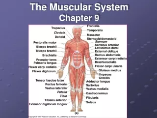
Major Muscles of the Body • Naming the Muscles • Students often learn the muscle names, locations, origins, insertions, and actions. • Muscle names are based on Latin word parts that relate to a fact about the muscle, such as: • Appearance • Location • Action • Relationship to other body parts. • Examples: pectoralis major, trapezius, biceps brachii