Figure 7.21a
Sacral promontory. Ala. Body of first sacral vertebra. Transverse ridges (sites of vertebral fusion). Anterior sacral foramina. Apex. Coccyx. (a) Anterior view. Figure 7.21a. Facet of superior articular process. Sacral canal. Body. Ala. Auricular surface. Median
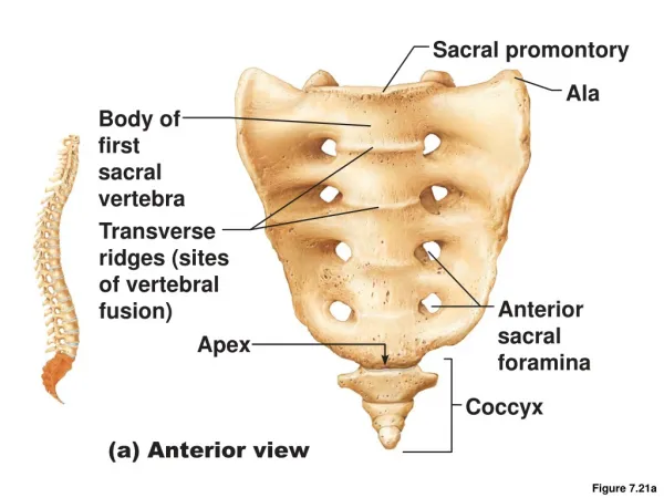

Figure 7.21a
E N D
Presentation Transcript
Sacral promontory Ala Body of first sacral vertebra Transverse ridges (sites of vertebral fusion) Anterior sacral foramina Apex Coccyx (a) Anterior view Figure 7.21a
Facet of superior articular process Sacral canal Body Ala Auricular surface Median sacral crest Lateral sacral crest Posterior sacral foramina Sacral hiatus Coccyx (b) Posterior view Figure 7.21b
Thoracic Cage • Composed of • Thoracic vertebrae • Sternum • Ribs and their costal cartilages • Functions • Protects vital organs of thoracic cavity • Supports shoulder girdle and upper limbs • Provides attachment sites for many muscles, including intercostal muscles used during breathing
Sternum (Breastbone) • Three fused bones • Manubrium • Articulates with clavicles and ribs 1 and 2 • Body • Articulates with costal cartilages of ribs 2 through 7 • Xiphoid process • Site of muscle attachment • Not ossified until ~ age 40
Ribs and Their Attachments • 12 pairs • All attach posteriorly to thoracic vertebrae • Pairs 1 through 7 • True (vertebrosternal) ribs • Attach directly to the sternum by individual costal cartilages
Ribs and Their Attachments • Pairs 8 through12 • False ribs • Pairs 8–10 also called vertebrochondral ribs • Attach indirectly to sternum by joining costal cartilage of rib above • Pairs 11–12 also called vertebral (floating) ribs • No attachment to sternum
Jugular notch Clavicular notch Manubrium Sternal angle Body Sternum True ribs (1–7) Xiphisternal joint Xiphoid process False ribs (8–12) Intercostal spaces Costal cartilage Costal margin L1 Vertebra Floating ribs (11, 12) (a) Skeleton of the thoracic cage, anterior view Figure 7.22a
Structure of a Typical Rib • Main parts: • Head • Articulates posteriorly with facets (demifacets) on bodies of two adjacent vertebrae • Neck • Tubercle • Articulates posteriorly with transverse costal facet of same-numbered thoracic vertebra • Shaft
Transverse costal facet (for tubercle of rib) Superior costal facet (for head of rib) Angle of rib Body of vertebra Head of rib Intervertebral disc Neck of rib Tubercle of rib Shaft Sternum Cross- section of rib Costal groove Costal cartilage (a) Vertebral and sternal articulations of atypical true rib Figure 7.23a
Articular facet on tubercle of rib Spinous process Shaft Transverse costal facet (for tubercle of rib) Ligaments Neck of rib Body of thoracic vertebra Head of rib Superior costal facet (for head of rib) (b) Superior view of the articulation between arib and a thoracic vertebra Figure 7.23b
Appendicular Skeleton • Bones of the limbs and their girdles • Pectoral girdle attaches the upper limbs to the body trunk • Pelvic girdle secures the lower limbs
Pectoral Girdle (Shoulder Girdle) • Clavicles and the scapulae • Attach the upper limbs to the axial skeleton • Provide attachment sites for muscles that move the upper limbs
Acromio- clavicular joint Clavicle Scapula (a) Articulated pectoral girdle Figure 7.24a
Clavicles (Collarbones) • Flattened acromial (lateral) end articulates with the scapula • Cone-shaped sternal (medial) end articulates with the sternum • Act as braces to hold the scapulae and arms out laterally
Sternal (medial) end Posterior Anterior Acromial (lateral) end (b) Right clavicle, superior view Figure 7.24b
Scapulae (Shoulder Blades) • Situated on the dorsal surface of rib cage, between ribs 2 and 7 • Flat and triangular, with three borders and three angles • Seven large fossae, named according to location
Suprascapular notch Acromion Superior border Coracoid process Superior angle Glenoid cavity Subscapular fossa Lateral border Medial border Inferior angle (a) Right scapula, anterior aspect Figure 7.25a
Coracoid process Suprascapular notch Superior angle Acromion Supraspinous fossa Glenoid cavity at lateral angle Spine Infraspinous fossa Medial border Lateral border (b) Right scapula, posterior aspect Figure 7.25b
Supraspinous fossa Supraglenoid tubercle Acromion Coracoid process Glenoid cavity Spine Supraspinous fossa Infraglenoid tubercle Infraspinous fossa Infraspinous fossa Subscapular fossa Subscapular fossa Posterior Anterior (c) Right scapula, lateral aspect Inferior angle Figure 7.25c
The Upper Limb • 30 bones form the skeletal framework of each upper limb • Arm • Humerus • Forearm • Radius and ulna • Hand • 8 carpal bones in the wrist • 5 metacarpal bones in the palm • 14 phalanges in the fingers
Humerus • Largest, longest bone of upper limb • Articulates superiorly with glenoid cavity of scapula • Articulates inferiorly with radius and ulna
Greater tubercle Head of humerus Lesser tubercle Anatomical neck Inter- tubercular sulcus Deltoid tuberosity Lateral supracondylar ridge Coronoid fossa Radial fossa Medial epicondyle Capitulum Trochlea (a) Anterior view Figure 7.26a
Bones of the Forearm • Ulna • Medial bone in forearm • Forms the major portion of the elbow joint with the humerus • Radius • Lateral bone in forearm • Head articulates with capitulum of humerus and with radial notch of ulna • Interosseous membrane connects the radius and ulna along their entire length
Radial notch of the ulna Olecranon process Trochlear notch Head Head of radius Coronoid process Neck Radial tuberosity Neck of radius Proximal radioulnar joint Interosseous membrane Ulna Radius Ulnar notch of the radius Radius Head of ulna Styloid process of ulna Styloid process of radius Distal radioulnar joint Styloid process of radius (a) Anterior view (b) Posterior view Figure 7.27a-b
Olecranon process View Trochlear notch Coronoid process Radial notch (c) Proximal portion of ulna, lateral view Ulnar notch of radius Articulation for lunate Articulation for scaphoid Styloid process Styloid process Head of ulna View (d) Distal ends of the radius and ulna at the wrist Figure 7.27c-d
Coronoid fossa Humerus Capitulum Medial epicondyle Trochlea Head of radius Coronoid process of ulna Radial tuberosity Radial notch Radius Ulna (c) Anterior view at the elbow region Olecranon fossa Humerus Olecranon process Lateral epicondyle Medial epicondyle Head Ulna Neck Radius (d) Posterior view of extended elbow Figure 7.26c-d
Hand: Carpus • Eight bones in two rows • Proximal row • Scaphoid, lunate, triquetrum, and pisiform proximally • Distal row • Trapezium, trapezoid, capitate, and hamate distally • Only scaphoid and lunate articulate with radius to form wrist joint
Hand: Metacarpus and Phalanges • Metacarpus • Five metacarpal bones (#1 to #5) form the palm • Phalanges • Each finger (digit), except the thumb, has three phalanges—distal, middle, and proximal • Fingers are numbered 1–5, beginning with the thumb (pollex) • Thumb has no middle phalanx
Phalanges • Distal • Middle • Proximal Metacarpals • Head • Shaft • Base Sesamoid bones Carpals Carpals Carpals • Trapezium • Hamate • Trapezium • Trapezoid • Capitate • Trapezoid • Scaphoid • Pisiform • Scaphoid • Triquetrum Radius • Lunate Ulna Radius (a) Anterior view of left hand (b) Posterior view of left hand Figure 7.28a-b
Pelvic (Hip) Girdle • Two hip bones (each also called coxal bone or os coxae) • Attach the lower limbs to the axial skeleton with strong ligaments • Transmit weight of upper body to lower limbs • Support pelvic organs • Each hip bone consists of three fused bones: ilium, ischium, and pubis • Together with the sacrum and the coccyx, these bones form the bony pelvis
Base of sacrum Iliac crest Sacroiliac joint Iliac fossa Anterior superior iliac spine Sacral promontory Coxal bone (os coxae or hip bone) Anterior inferior iliac spine llium Sacrum Pubic bone Pelvic brim Coccyx Acetabulum Pubic tubercle Ischium Pubic crest Pubic symphysis Pubic arch PLAY Animation: Rotatable pelvis Figure 7.29
Hip Bone • Three regions • Ilium • Superior region of the coxal bone • Auricular surface articulates with the sacrum (sacroiliac joint) • Ischium • Posteroinferior part of hip bone • Pubis • Anterior portion of hip bone • Midline pubic symphysis joint
Anterior gluteal line Ilium Ala Posterior gluteal line Iliac crest Posterior superior iIiac spine Anterior superior iliac spine Posterior inferior iliac spine Inferior gluteal line Greater sciatic notch Anterior inferior iliac spine Ischial body Acetabulum Ischial spine Pubic body Lesser sciatic notch Pubis Ischium Inferior ramus of pubis Ischial tuberosity Obturator foramen Ischial ramus (a) Lateral view, right hip bone Figure 7.30a
Ilium Iliac fossa Iliac crest Posterior superior iliac spine Anterior superior iliac spine Posterior inferior iliac spine Anterior inferior iliac spine Auricular surface Body of the ilium Arcuate line Greater sciatic notch Superior ramus of pubis Ischial spine Lesser sciatic notch Pubic tubercle Obturator foramen Articular surface of pubis (at pubic symphysis) Ischium Ischial ramus Inferior ramus of pubis (b) Medial view, right hip bone Figure 7.30b
Comparison of Male and Female Pelves • Female pelvis • Adapted for childbearing • True pelvis (inferior to pelvic brim) defines birth canal • Cavity of the true pelvis is broad, shallow, and has greater capacity
Comparison of Male and Female Pelves • Male pelvis • Tilted less forward • Adapted for support of male’s heavier build and stronger muscles • Cavity of true pelvis is narrow and deep
The Lower Limb • Carries the weight of the body • Subjected to exceptional forces • Three segments of the lower limb • Thigh: femur • Leg: tibia and fibula • Foot: 7 tarsal bones in the ankle, 5 metatarsal bones in the metatarsus, and 14 phalanges in the toes
Femur • Largest and strongest bone in the body • Articulates proximally with the acetabulum of the hip and distally with the tibia and patella
Neck Fovea capitis Greater trochanter Head Inter- trochanteric crest Lesser trochanter Intertrochanteric line Gluteal tuberosity Linea aspera Apex Anterior Facet for lateral condyle of femur Facet for medial condyle of femur Lateral condyle Medial and lateral supra- condylar lines Lateral epicondyle Surface for patellar ligament Intercondylar fossa Medial condyle Posterior Adductor tubercle (a) Patella (kneecap) Lateral epicondyle Medial epicondyle Patellar surface Anterior view Posterior view (b) Femur (thigh bone) Figure 7.31
Bones of the Leg • Tibia • Medial leg bone • Receives the weight of the body from the femur and transmits it to the foot • Fibula • Not weight bearing; no articulation with femur • Site of muscle attachment • Connected to tibia by interosseous membrane • Articulates with tibia via proximal and distal tibiofibular joints
Lateral condyle Intercondylar eminence Head Medial condyle Proximal tibiofibular joint Tibial tuberosity Interosseous membrane Anterior border Fibula Tibia Distal tibiofibular joint Articular surface Lateral malleolus Medial malleolus (a) Anterior view Figure 7.32a
Articular surface of medial condyle Articular surface of lateral condyle Medial condyle Head of fibula Interosseous membrane Tibia Fibula Articular surface Medial malleolus Lateral malleolus (b) Posterior view Figure 7.32b