Download
1 / 76
760 likes | 1.2k Views
Explore the close relationship and functions of the stomach, pancreas, and intestines, and learn about the phases of digestion. Discover the role of the laboratory in investigating gastrointestinal conditions, including diseases like Helicobacter pylori infections.

E N D
Introduction • The stomach, intestinal tract, and pancreas are closely related, both anatomically and functionally, and clinical manifestations, such as diarrhea or malabsorption, may be associated with disease or disorders of any of these organs • Advances in imaging techniques and improvements in endoscopic procedures has led to many traditional laboratory tests of gastrointestinal (GI) and pancreatic function becoming obsolete. • However, in recent years, there has been a resurgence in the role of the laboratory in the investigation of the GI tract, particularly with the development of noninvasive biomarkers of GI tract inflammation and in the detection of pancreatic insufficiency.
PHASES OF DIGESTION • The processes of digestion can be divided into neurogenic, gastric, and intestinal phases. • Neurogenic Phase • The neurogenic, or cephalic, phase is initiated by the intake of food into the mouth; the sight, smell, and taste of food stimulates the cerebral cortex and subsequently the vagal nuclei.
PHASES OF DIGESTION • Gastric Phase • When food enters the stomach the resulting distention initiates the gastric phase of digestion. • Hydrochloric acid release is caused by direct vagal stimulation of the parietal cells, local distention of the antrum, and vagal stimulation of antral cells to secrete gastrin. • Gastrin also stimulates antral motility, secretion of pepsinogens and of pancreatic fluid rich in enzymes, and release of hormones such as secretin, insulin, acetylcholine, somatostatin, and pancreatic polypeptide.
PHASES OF DIGESTION • As a result of the acid environment pepsinogen is rapidly converted to the active proteolytic enzyme pepsin. • Food is mixed by contractions of the stomach and is partially degraded into chyme by the chemical secretions of the stomach. • The pylorus plays a role in emptying chyme into the duodenum by virtue of its strong musculature.
PHASES OF DIGESTION • Intestinal Phase • The intestinal phase of digestion begins when the weakly acidic digestive products of proteins enter the duodenum. • Many hormones and other regulatory peptides are released by both neural and local stimulation and act within the GI tract to regulate digestion and absorption. • Digestion, absorption, and storage functions are stimulated or inhibited by different hormones, creating a control system that regulates the action of intestinal hormones and provides for secretion of bile acids, bicarbonate, and numerous enzymes involved in the digestion of food.
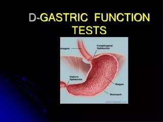
STOMACH: DISEASES ANDLABORATORY INVESTIGATIONS • Growth in endoscopic procedures, with direct visualization of the interior of the stomach, has largely removed the need for the laboratory to carry out investigation of gastric contents. • Situations remain, however, in which the laboratory continues to play a role in diagnosing gastric diseases and in monitoring the effectiveness of treatment.
STOMACH: DISEASES AND LABORATORY INVESTIGATIONSHelicobacter pylori • H. pylori is the predominant cause of gastric and duodenal ulcers, the remainder being associated with the long-term use of nonsteroidal anti-inflammatory drugs (NSAIDS) and, rarely, gastrinomas. • H. pylori is present in the mucus layer of the stomach in half of the world’s population. • In Europe 30% to 50% of adults and in the United States at least 20% of the adult population are infected with the bacterium. • Chronic infection produces an inflammatory response (gastritis) and carries an increased risk for developing a peptic ulcer (3- to 10-fold) and/or adenocarcinoma (2- to 10-fold).
STOMACH: DISEASES AND LABORATORY INVESTIGATIONSHelicobacter pylori • Up to 90% of gastric cancer patients are infected with H. pylori. • In a European study comparing the prevalence of H. pylori versus gastric cancer rates in 13 countries, a significant correlation was found between the infection rate and gastric cancer incidence and mortality. • H. pylori may cause dyspepsia in the absence of an ulcer, and current recommendations suggest a low threshold for testing for H. pylori and some advocate treatment without testing.
STOMACH: DISEASES AND LABORATORY INVESTIGATIONSHelicobacter pylori • The mode of transmission of H. pylori is unclear. • In many cases the infection appears to originate in childhood, presumably by the fecal-oral route, because the prevalence is higher in developing countries and is inversely related to food hygiene. • Almost all individuals infected with H. pylori develop chronic gastritis, but only 10% of cases manifest as peptic ulcers. • H. pylori infection predominantly affects the gastric mucosa, with the antrum usually the most densely colonized area. • At least 95% of patients with duodenal ulcers have H. pylori infection, and eradication of the organism results in healing of the ulcer and a reduction in relapse rates.
STOMACH: DISEASES AND LABORATORY INVESTIGATIONSHelicobacter pylori • This variation is governed by a number of factors including the site of infection, virulence factors (eg, vacuolating cytotoxins [VAC], CagA protein), mucus secretion and extent of pepsinogen secretion. • Infection of the mid-body of the stomach is the commonest form, occurs in people with a strong immune system and involves a type of H. pylori with low expression of CagA and VAC. • However, if the infection is in the antrum, the inflammation causes the G cells to become hyperactive with a resulting disproportionate secretion of gastrin in response to food and gastric distention and consequent increases in acid output.
STOMACH: DISEASES AND LABORATORY INVESTIGATIONSHelicobacter pylori • Basal acid output has been shown to be higher in H. pylori–infected subjects, and this resolves after eradication of the organism. • Hypergastrinemia is believed to be only one of the mechanisms leading to increased acid output. • H. pylori produces urease, and hydrolysis of this endogenously produced urea to bicarbonate and ammonia may create a more hospitable environment for the survival of the organism in the stomach. • This ability of H. pylori to hydrolyze urea forms the basis of urea breath tests and direct urease tests on gastric biopsy samples.
STOMACH: DISEASES AND LABORATORY INVESTIGATIONSHelicobacter pylori : Diagnostic Tests for H. pylori • Numerous invasive and noninvasive diagnostic tests for H. pylori have been described: • Invasive tests: Using gastric mucosal biopsy samples • Histology: Microscopy after Giemsa, silver staining or IHC staining • Direct urease test: Biopsy included in urea/ indicator solution—visual end point • Culture: Incubation in suitable media for 4 to 10 days • Polymerase chain reaction: Amplification of specific DNA sequences Direct urease test
STOMACH: DISEASES AND LABORATORY INVESTIGATIONSHelicobacter pylori : Diagnostic Tests for H. pylori • Noninvasive tests: Using breath, blood, saliva, or feces • Breath tests: Rise in 14CO2 or 13CO2 after ingestion of 14C- or 13C-labeled urea • The breath test has sensitivity and specificity for H. pylori in excess of 95% and can be used both for diagnosis and to assess the success of eradication therapy.
STOMACH: DISEASES AND LABORATORY INVESTIGATIONSHelicobacter pylori : Diagnostic Tests for H. pylori Serum, saliva, or feces tests • Serologic methods are available to detect H. pylori–specific antibodies (IgG or IgA), but have some drawbacks compared to the urea breath test. • The systemic antibody response is variable, with equivocal results often occurring in subjects older than 50 years. • The sensitivity (92%) and specificity (83%) are also lower than those for the breath test. • Serologic tests cannot be used to confirm eradication of the bacterium because of the persistence of the antibodies for variable periods after completion of treatment.
STOMACH: DISEASES AND LABORATORY INVESTIGATIONSHelicobacter pylori : Diagnostic Tests for H. pylori • H. pylori is shed in feces, and several tests have been described that can detect the organism. • Polyclonal or monoclonal antibodies to H. pylori can be configured into various immunoassay formats, although sensitivity and specificity are lower than for breath tests. • Commercial kits are available that use polymerase chain reaction to amplify nuclear sequences specific for H. pylori in feces (or saliva) and have a sensitivity and specificity of 95% and 94%, respectively.
STOMACH: DISEASES AND LABORATORY INVESTIGATIONS Gastric Acid Secretion and Gastrinomas • Collection of gastric juice and analysis of acid output was at one time extensively carried out in the investigation of possible gastrinomas. • This invasive technique has now been replaced by the greater availability of: • plasma gastrin measurements, • endoscopy, and • imaging modalities, including: • computed tomography (CT) and magnetic resonance imaging (MRI).
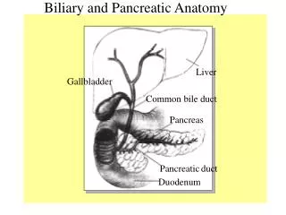
STOMACH: DISEASES AND LABORATORY INVESTIGATIONS Gastric Acid Secretion and Gastrinomas: Gastrin • Three molecular forms of gastrin occur in blood and tissues: G-34, G-17, and G-14. • Gastrin is produced and stored mainly by: • endocrine cells (G cells) of the antral mucosa • to a lesser extent by G cells of the proximal duodenum and • Δ cells of the pancreatic islets Proximal duodenum
STOMACH: DISEASES AND LABORATORY INVESTIGATIONS Gastric Acid Secretion and Gastrinomas: Gastrin • After secretion, gastrin is transported by the blood through the liver to the parietal cells of the fundus of the stomach, where it stimulates the secretion of gastric acid. • Gastrin is secreted in response to antral distention from food and by the presence of amino acids, peptides, and polypeptides in the stomach from partially digested proteins. • Other stimuli of gastrin include alcohol, caffeine, insulin induced hypoglycemia, and calcium.
STOMACH: DISEASES AND LABORATORY INVESTIGATIONS Gastric Acid Secretion and Gastrinomas: Gastrin • Maximal secretion of gastrin occurs at an antral pH of 5 to 7. • At pH 2.5, secretion of gastrin is reduced by approximately 80%, with maximal suppression occurring at pH 1. • Secretion is inhibited by the direct action of acid on the G cells. • This negative feedback prevents excess acid production regardless of the stimulant.
STOMACH: DISEASES AND LABORATORY INVESTIGATIONS Gastric Acid Secretion and Gastrinomas: Gastrin • The principal circulating form of gastrin is in healthy individuals and in patients with hypergastrinemia is G-34. • Trypsin cleaves G-34 into two fragments one of which has the amino acid sequence of G-17. • On a molar basis, G-17 is 6 to 8 times more potent than G-34 as a stimulant of gastric acid secretion. • In the fasting state, the ratio of G-34 to G-17 is approximately 2:1. • After meals, the concentration of G-34 doubles but that of G-17 increases fourfold so the ratio approaches 1:1.
STOMACH: DISEASES AND LABORATORY INVESTIGATIONS Gastric Acid Secretion and Gastrinomas: Gastrinoma & the Zollinger-Ellison Syndrome • In 1955, Zollinger and Ellison described a syndrome consisting of: • Multiple peptic ulcers, • Gastric acid hypersecretion, and • Non–β islet cell tumors of the pancreas secreting gastrin. • Gastrinomas are a rare cause (<0.5%) of gastroduodenal ulcers. • The persistently high circulating gastrin concentrations lead to hypersecretion of gastric acid and increased parietal cell mass.
STOMACH: DISEASES AND LABORATORY INVESTIGATIONS Gastric Acid Secretion and Gastrinomas: Gastrinoma & the Zollinger-Ellison Syndrome • Duodenal ulceration and ulcers resistant to standard therapies must be considered suspicious. • Gastrinomas are most often sporadic, but also may be associated with multiple endocrine neoplasia type 1 (MEN1, Wermer’s syndrome), a syndrome characterized by the presence of two or more tumors sited in the pituitary, parathyroid glands, or pancreas. • MEN1 is also associated with an increased prevalence of adrenal and thyroid.
STOMACH: DISEASES AND LABORATORY INVESTIGATIONS Gastric Acid Secretion and Gastrinomas: Gastrinoma & the Zollinger-Ellison Syndrome • Gastrinomas are commonly located in the pancreas, but can arise from the stomach, duodenum, or other tissues. • They are more often (60%) malignant than benign, with metastases frequently present at the time of diagnosis. • Measurement of plasma gastrin in a fasting sample is the initial step in aiding the differential diagnosis (concentration more than 10 times the upper limit of the reference range). • A mildly elevated plasma gastrin concentration may be observed in long-term PPI therapy, hypochlorhydria, pernicious anemia, and G-cell hyperplasia.
INTESTINAL DISORDERS & THEIR LABORATORY INVESTIGATIONCeliac Disease: Pathophysiology of Celiac Disease • Celiac disease occurs in genetically predisposed subjects as a consequence of an inappropriate T cell–mediated immune response to ingestion of gluten from wheat and to similar proteins in barley and rye. • The role of genetic factors in celiac disease has been recognized for many years; a 70% concordance for celiac disease has been reported in identical twins • The major genetic component has been localized to the human leukocyte antigen (HLA) region of chromosome 6. • Approximately 95% of subjects with celiac disease express a specific HLA heterodimer (HLA DQ2 α/β).
INTESTINAL DISORDERS & THEIR LABORATORY INVESTIGATIONCeliac Disease: Pathophysiology of Celiac Disease • The external trigger to the development of celiac disease in genetically susceptible individuals is found in gluten, which is the complex group of proteins found in wheat and other grains. • The major toxic proteins of wheat are the gliadins that account for 50% of the wheat protein, with homologous proteins (the hordeins and secalins) occurring in barley and rye, respectively. • All proteins (and peptides) that are toxic to the small bowel mucosa in subjects with celiac disease contain large amounts of glutamine.
INTESTINAL DISORDERS & THEIR LABORATORY INVESTIGATIONCeliac Disease: Pathophysiology of Celiac Disease • The identification in 1997 of small bowel tissue transglutaminase as the autoantigen of celiac disease has led to a greater understanding of the pathogenesis of the condition. • The tissue transglutaminases are a family of calcium-dependent enzymes that are released from cells during wound healing. • They catalyze the cross-linking or deamidation of proteins leading to stabilization of the wound area. • Expression of the enzyme is increased during apoptosis and in active celiac disease. • Deamidation of gliadin peptides enhances their binding to HLA DQ2/DQ8 and increases recognition of these peptides by gut-derived T cells from subjects with celiac disease.
INTESTINAL DISORDERS & THEIR LABORATORY INVESTIGATIONCeliac Disease: Pathophysiology of Celiac Disease
INTESTINAL DISORDERS & THEIR LABORATORY INVESTIGATIONCeliac Disease: Pathophysiology of Celiac Disease • The characteristic enteropathy is then induced by the release of interferon-γ and other proinflammatory cytokines. • A 33–amino acid peptide of gluten appears to be the primary initiator of the inflammatory response. • It is resistant to breakdown by all gastric, pancreatic, and brush border membrane proteases, thus allowing it to reach the small intestine intact. • Homologs of the peptide are found in food grains that are toxic to patients with celiac disease but are absent from nontoxic food grains.
INTESTINAL DISORDERS & THEIR LABORATORY INVESTIGATIONCeliac Disease: Pathophysiology of Celiac Disease • Increased intestinal permeability in untreated celiac disease that is reversible on withdrawal of gluten from the diet has been recognized since the early 1980s. • Evidence suggests that this increase in paracellular permeability may be mediated by increased expression of zonulin, modulator of intercellular tight junctions (TJ) (a protein that opens small intestinal TJ), or by decreased expression of intercellular epithelial cell adhesion molecules such as catenin, and cadherin. • The zonulin pathway is now thought to play a significant role in the entry of allergens into cells and hence in the autoimmune response.
INTESTINAL DISORDERS & THEIR LABORATORY INVESTIGATIONCeliac Disease: Pathophysiology of Celiac Disease
INTESTINAL DISORDERS & THEIR LABORATORY INVESTIGATIONCeliac Disease: Tests for Celiac Disease • Serologic tests have played a significant role in raising awareness of the high prevalence of this disorder and appropriately standardized tests have high clinical sensitivity and specificity for diagnosis and monitoring of compliance with a gluten free diet.
INTESTINAL DISORDERS & THEIR LABORATORY INVESTIGATION Bacterial Overgrowth • The proximal small intestine (duodenum and jejunum) normally contains few bacteria. • Most ingested bacteria do not survive the acidic environment of the stomach; therefore few live organisms enter the small bowel. • The motility of the jejunum prevents fecal-type organisms from progressing up into the jejunum from the cecum. • The ileum normally contains some fecal-type bacteria. • Colonization of the upper small bowel is described as bacterial overgrowth and usually occurs as a consequence of other abnormalities (structural or motility disorders) of the small intestine.
INTESTINAL DISORDERS & THEIR LABORATORY INVESTIGATION Bacterial Overgrowth • Gastric hypochlorhydria (associated with the use of PPIs) may contribute to bacterial overgrowth. • Abnormalities that can result in bacterial overgrowth include: • The systemic immune system, eg, isolated IgA deficiency, hypogammaglobulinemia, combined immunodeficiency, infection with the human immunodeficiency virus [HIV] • Impaired motility, pancreatic insufficiency, intrinsic small bowel disease, or surgically induced blind loops are often associated with bacterial overgrowth.
INTESTINAL DISORDERS & THEIR LABORATORY INVESTIGATION Bacterial Overgrowth • Small bowel bacterial overgrowth can be asymptomatic or associated with nonspecific symptoms that include abdominal distention and diarrhea resembling IBS. • Bacteria colonizing the small bowel (eg, Escherichia • coli and Bacteroides species) deconjugate and dehydroxylate bile salts, resulting in conjugated bile salt deficiency and mild fat malabsorption. • Bacterial metabolism of vitamin B12 can occur, leading to deficiency, whereas plasma folate concentration may be increased owing to production by the bacteria.
INTESTINAL DISORDERS & THEIR LABORATORY INVESTIGATION Bacterial Overgrowth • The gold standard diagnostic procedure is microbiologic examination of small bowel contents, but this is seldom practical. • Noninvasive procedures based on oral administration of substances metabolized by bacteria to yield products that can be detected in breath are now the most common tests for bacterial overgrowth.
INTESTINAL DISORDERS & THEIR LABORATORY INVESTIGATION Bacterial Overgrowth • Small bowel bacterial breath tests in routine use involve test substances labeled with 13C and the breath hydrogen tests. • Lactulose is not normally absorbed from the small bowel and is therefore available for metabolism by bacteria throughout the gut; alternatively, labeled glucose can be used. • In a normal subject, breath hydrogen does not increase until the substrate reaches the large intestine and the time from ingestion to a rise in breath hydrogen is an indicator of small bowel transit time. • In bacterial overgrowth an early rise in breath hydrogen of at least 20 ppm (20 μl/L) is observed within 30 minutes of ingestion.
INTESTINAL DISORDERS & THEIR LABORATORY INVESTIGATION Bile Acid Malabsorption • Bile acids are synthesized in the liver and pass into the lumen of the small bowel via the gallbladder. • Bile acids are present in bile as taurine or glycine conjugates; because the pH of bile is slightly alkaline and contains significant amounts of sodium and potassium, most of the bile acids and their conjugates exist as salts (ie, bile salts). • In practice, the terms bile acids and bile salts are frequently used synonymously. • Their major function is to act as surface-active agents, forming micelles and facilitating the digestion of triglycerides and the absorption of cholesterol and fat-soluble vitamins.
INTESTINAL DISORDERS & THEIR LABORATORY INVESTIGATION Bile Acid Malabsorption • Little reabsorption of bile acids occurs in the proximal small bowel, but normally more than 90% is reabsorbed in the terminal ileum. • The bile acids return to the liver via the portal circulation and can be resecreted into bile (enterohepatic circulation). • Less than 10% of secreted bile acid is lost in the feces (~0.2 to 0.6 g/24 h). • Bile acid malabsorption leading to chronic diarrhea occurs when ileal disease is present, or after resection of the terminal ileum; it also may occur after cholecystectomy and is present in some patients with IBS.
INTESTINAL DISORDERS & THEIR LABORATORY INVESTIGATION Bile Acid Malabsorption • Malabsorption of bile salts produces diarrhea by two different mechanisms. • When significant bile salt depletion occurs, the deficiency of intraluminal bile salts leads to fat malabsorption and steatorrhea. • More commonly, malabsorption of bile salts in the ileum leads to increased concentrations of bile salts in the colon, where they alter water and electrolyte absorption. • This leads to net secretion of water into the lumen and diarrhea. • Bile salt malabsorption is probably an underdiagnosed condition and should be suspected in patients with unexplained chronic diarrhea.
INTESTINAL DISORDERS & THEIR LABORATORY INVESTIGATION Bile Acid Malabsorption • Two blood tests have been proposed: fibroblast growth factor-19 (FGF-19) and 7α-hydroxy-4-cholesten-3-one (C4). • C4 is the intermediary step between cholesterol and taurocholic acid and reflects the activity of hepatic cholesterol 7α-hydroxylase and therefore, because this is the rate-limiting enzyme, the rate of bile acid synthesis. • Bile acid malabsorption is associated with increased plasma concentrations of C4 as hepatic synthesis is up-regulated to maintain the pool of circulating bile acids.
INTESTINAL DISORDERS & THEIR LABORATORY INVESTIGATION Bile Acid Malabsorption • The presence of bile acids in the ileum has been shown to stimulate the production of FGF-19. • Good correlation has been demonstrated between both serum C4 and FGF-19. • Several publications have demonstrated good sensitivity for identifying bile acid–induced diarrhea. • With growing awareness of the role of bile salt malabsorption in chronic diarrhea in a proportion of patients with IBS or inflammatory bowel disease (IBD), and the therapeutic effectiveness of cholestyramine, measurement of C4 and/or FGF-19 may become routine.
INTESTINAL DISORDERS & THEIR LABORATORY INVESTIGATION Inflammatory Bowel Disease and Irritable Bowel Syndrome • Intestinal symptoms including abdominal pain or discomfort with diarrhea or constipation are common to both IBD and IBS. • IBD includes ulcerative colitis (UC) and Crohn disease (CD), as well as a number of microscopic inflammatory bowel conditions. • IBS is a functional bowel disorder for which there is no identifiable pathologic process or known cause. • Although IBS may produce symptoms that are sufficient to interfere with daily life, it is seldom associated with serious morbidity.
INTESTINAL DISORDERS & THEIR LABORATORY INVESTIGATION Inflammatory Bowel Disease (IBD) and Irritable Bowel Syndrome (IBS) • IBS may affect up to 10% to 20% of the adult population in the developed world, with a typical age at presentation of 20 to 30 years and a female preponderance. • Both UC and CD are chronic conditions that tend to follow a remitting and relapsing course. • The prognosis of patients with CD tends to be worse than that of UC, although in the latter it is estimated that up to 10% of patients will require surgical intervention within 10 years of diagnosis.
INTESTINAL DISORDERS & THEIR LABORATORY INVESTIGATION Inflammatory Bowel Disease (IBD) and Irritable Bowel Syndrome (IBS) • The cause of IBD is not fully understood, but both genetic and environmental factors have been implicated. • Genomewide association studies have identified over 160 susceptibility loci/genes that are significantly associated with IBD. • Studies in monozygotic twins, however, have shown a lower than expected concordance rate (15% to 19% for UC, 27% to 50% for CD) if IBD were simply an inherited disorder.
INTESTINAL DISORDERS & THEIR LABORATORY INVESTIGATION Inflammatory Bowel Disease (IBD) and Irritable Bowel Syndrome (IBS) • The enteric GI microbiota are now accepted as a central etiologic factor in the pathogenesis of IBD, although the mechanistic details are lacking. • Tissue damage is a consequence of neutrophil activation and the production of proinflammatory cytokines and chemokines. • The success of treatments based on immunosuppressive agents or tumor necrosis factor-alpha (TNF-α) inhibitors appears to support an immune basis for the development of IBD. • Environmental factors implicated in triggering the development of active disease include a protective effect of breastfeeding and an increased risk in patients who have recurrent GI infections or exposure to drug therapy (antibiotics, oral contraceptives, NSAIDs) along with socioeconomic factors, stress, smoking, and diet.
INTESTINAL DISORDERS & THEIR LABORATORY INVESTIGATION Inflammatory Bowel Disease (IBD) and Irritable Bowel Syndrome (IBS) • The conventional diagnostic pathway includes a full blood count to detect/exclude anemia, markers of generalized inflammation (erythrocyte sedimentation rate [ESR] and C-reactive protein [CRP]) and serologic testing for celiac disease. • In the past 20 years, there have been significant advances in biomarkers that can aid in differentiating IBD from IBS and in monitoring disease activity or response to treatment. • Most of these are fecal markers.