Download

1 / 49
700 likes | 2.01k Views
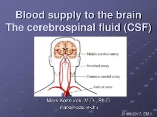
BLOOD SUPPLY OF BRAIN. BY: DR. IBRAR AHMED HASHMI. ARTERIAL SUPPLY OF BRAIN. COMMON CAROTID ARTERY 70% blood is delivered to ICA

E N D
BLOOD SUPPLY OF BRAIN BY: DR. IBRAR AHMED HASHMI
ARTERIAL SUPPLY OF BRAIN COMMON CAROTID ARTERY • 70% blood is delivered to ICA • Carotid bifurcation is a physiological stenosis due to inertial forces of blood flow divert main flow stream from midvessel to a path along vessel margin at flow divider • CCA divides lateral to upper border of thyriod cartilage: C3-4 intervertebral disc. • ECA arises anterior and medial to ICA(95%)
EXTERNAL CAROTID ARTERY • ASCENDING PHARYNGEAL ARTERY • SUPERIOR THYROID ARTERY • LINGUAL ARTERY • EXTERNAL MAXILLARY=FACIAL ARTERY • OCCIPITAL ARTERY • POSTERIOR AURICULAR ARTERY • SUPERFICIAL TEMPORAL ARTERY • INTERNAL MAXILLARY ARTERY
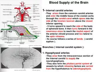
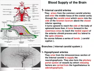
INTERNAL CAROTID ARTERY • CERVICAL SEGMENT • PETROUS SEGMENT • CAVERNOUS SEGMENT • SUPRACLINOID SEGMENT
INTERNAL CAROTID ARTERY • CERVICAL SEGMENT: • Ascends posterior and medial to ECA: enters carotid canal of petrous bone; • NO BRANCHES • CAROTID BULB= CAROTID SINUS • Dilated proximal part of ICA with thinner media and thicker adventitia containing many receptor endings of glossopharyngeal nerve. • Baroreceptor responsive to change in arterial BP. • Hypersensitive carotid sinus: slight touch or neck movement initiates drop in BP and SA/AV blocks.
PETROUS SEGMENT: • Ascends briefly in carotid canal ; bends anteromedially in horizontal course(ant to tympanic cavity and cochlea); exits near post portion foramen lacerum;ascends to juxtasellar location and pierces dural layer of cavernous sinus. • BRANCHES: (rarely seen on angiograms) • CORTICOTYMPANIC A. • PTERYGOID (VIDIAN) A.(inconstant)
CAVERNOUS SEGMENT: • Ascends to posterior clinoid process, then turns anteriorly and superomedially through cavernous sinus: exits medial to ant clinoid process piercing dura. • BRANCHES: • Meningohypophysial trunk • Anterior meningeal artery • Cavernous rami for trigeminal ganglia, cavernous sinus and inf. petrosal sinuses.
SUPRACLINOID SEGMENT: • Ascends posterior + lateral b/w oculomotor and optic nerv. • BRANCHES: • OPHTHALMIC A. • SUPERIOR HYPOPHYSEAL A. (not routinely visualized) • PCOM • ANTERIOR CHOROIDAL A. • MCA • ACA
CAROTID SIPHON: (3rd + 4th part of ICA) • FLOW DIRECTION: C4---C1 • C4 SEGMENT= Before origin of ophthalmic a. • C3 SEGMENT= Genu of ICA. • C2 SEGMENT= Supraclinoid segment after origin of ophthalmic a. • C1 SEGMENT= Terminal segment of ICA b/w pCom + ACA.
ANTERIOR CEREBRAL ARTERY • A1 SEGMENT= HORIZONTAL PORTION b/w origin and aCom. • Inferior branches to optic nerve and chiasma • Superior branches to ant hypothalamus, septum pellucidum, ant commisure, fornix, columns, medial lenticulostriate artery to anteroinferior portion of corpus striatum. • A2 SEGMENT= INTERHEMISPHERIC PORTION after the origin of aCom.
BRANCHES: • Medial orbitofrontal artery. • Frontopolar artery. • Callosomarginal artery. • Pericallosal artery. • SUPPLY: anterior 2/3 of medial cerebral surface and 1cm of superomedial brain over convexity.
MIDDLE CEREBRAL ARTERY • Largest branch of ICA, arises lat to optic chiasma, passes horizontal and lateral direction to enter in sylvian fissure and divides into 2/3/4 branches • SUPPLY: • Lateral cerebrum • Insula • Anterior and Lateral temporal lobes • M1 SEGMENT: • Origin to MCA bifurcation • Lateral lenticulostriate • M2 SEGMENT: • Insular branches • M3 SEGMENT: • MCA branches beyond sylvian fissure
VERTEBRAL ARTERY • 1st branch of subclavian(95%) • Left vertebral arises directly from aortic arch in 5%. • Left artery is dominant in 50%, in 25% co dominant, in 25% right is dominant.
VERTEBRAL ARTERY • PREVERTEBRAL SEGMENT: Enters transverse foramina at C6, only muscular branches. • CERVICAL SEGMENT: Anterior meningeal artery. • ATLANTIC SEGMENT: exits through transverse foramina of atlas till it peierces dura to enter cranial cavity. Branch: Post. Meningeal. • INTRACRANIAL SEGMENT:
INTRACRANIAL SEGMENT • Ascends anteriorly + laterally around medulla to reach midline at pontomedullary junction and forms basilar artery with other vertebral a at clivus. • BRANCHES: • ANTERIOR + POSTERIOR SPINAL A. • PICA • BASILAR ARTERY BRANCHES: • AICA • INTERNAL AUDITORY A. • SUPERIOR CEREBELLAR A. • POSTERIOR CEREBRAL A. • MEDULLARY AND PONTINE PERFORATING ARTERIES
POSTERIOR CEREBRAL ARTERY • Originates from bifurcation of basilar artery, within interpeduncular cistern (in 15% as direct continuation of pCom) lies above occulomotor nerve and circles midbrain above tentorium. • P1 SEGMENT: • Origin to PCOM. • Posterior thalamoperforators • P2 SEGMENT: • Distal to PCOM • Thalamogeniculate • Posterior choroidal arteries. • TERMINAL CORTICAL BRANCHES.
ARTERIAL ANASTOMOSES OF BRAIN • AT BASE OF BRAIN: • CIRCLE OF WILLIS • DEVELOPMENTAL ANOMALIES: (3 transient carotid-basilar anastomoses appear in fetal life) • Primitive hypoglossal artery • Primitive acoustic artery • Persistent primitive trigeminal artery
CIRCLE OF WILLIS • Complete in 25%, incomplete in 75%. • Made by • Supraclinoid ICAs • A1 segment of ACA • ACOMs • PCOMs • P1 segment of PCAs
ARTERIAL ANASTOMOSES OF BRAIN • VIA SURFACE VESSELS: • LEPTOMENINGEAL ANASTOMOSES OF CEREBRUM: ACA MCA PCA • OF CEREBELLUM: SUP CEREBELLAR AICA PICA. • RETE MIRABILE: • ECA MIDDLE MENINGEAL/SUP TEMPORAL A. LEPTOMENINGEAL ACA/MCA.
NORMAL VARIANTS OF VASCULAR ANATOMY • ICA: • PERSISTENT EMBRYONIC ARTERIES • ABERRANT PETROUS PART ICA: Courses posterolateral • ECA: • MIDDLE MENINGEAL FROM OPHTHALMIC • VARIATION IN ORDER OF BRANCHING. • CIRCLE OF WILLIS: • HYPOPLASTIC PCOM • HYPOPLASTIC OR ABSENT A1 SEGMENT • FETAL PCA(FROM ICA) WITH ATRETIC P1 SEGMENT • HYPOPLASTIC ACOM.