Download

1 / 40
430 likes | 703 Views
Lab 1 Histology. Scott.lehbauer@lethbridgecollege.ab.ca. Introduction. Tissues - are groups of similar cells that are specialized to perform specific functions. For example, nervous tissue is specialized to conduct impulses that help control and coordinate body activities.

E N D
Lab 1 Histology Scott.lehbauer@lethbridgecollege.ab.ca
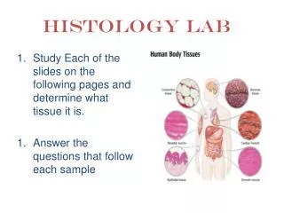
Introduction • Tissues - are groups of similar cells that are specialized to perform specific functions. For example, nervous tissue is specialized to conduct impulses that help control and coordinate body activities. • The study of tissues is called histology.
Epithelial Tissues • Are tissues that cover all the body surfaces inside and out. • are tissues that are composed of at least one layer of cells. • more specifically they make up the outer layer of the skin, form the inner lining of body cavities, cover organs, line the chambers inside hollow organs, and are the major tissue component of glands.
Epithelial tissues always have one side exposed to the outside or to an open space internally. • The underside of all epithelial tissues is anchored to connective tissue by a non-living layer called a basement membrane. Basement Membrane
Epithelial tissues are avascular (lack blood vessels) • Epithelial tissues obtain nutrients from the vessels in the underlying connective tissue. • Epithelial tissues reproduce rapidly, therefore injuries to epithelial tissues repair quickly.
Epithelial tissues are tightly packed together. (little intercellular space) • Epithelial tissues are classified according to their shape, arrangement and function of their cells.
Epithelial Tissue Classification • According to arrangement • 2. According to shape
According to Arrangement • Simple – one layer thick Simple Squamous Epithelium Simple Cuboidal Epithelium • Stratified – two or more layers Stratified Squamous Epithelium Transitional Epithelium
According to Shape Examples of Shapes of Epithelial Tissues. Wikipedia 2007.
Glandular – in skin, intestines, endocrine glands they make and secrete substances. • Absorption – through microvilli on the top surface of the cells. • Protection – the densely packed cells provide a protective layer for the underlying tissues. • Secretion – of fluid, enzymes mucus and other substances.
Simple Squamous Epithelium Description – one cell layer thick. • Squamous (thin) – thin cells that look like fried eggs.
Simple Squamous Epithelium Function – diffusion of gases • secrete serous fluid to lubricate covering of organs.
Simple Sqamous Epithelium Location • Lining of alveoli • Lining of capillaries • Lining of lymph vessels • Covering around organs
Simple Columnar Epithelium Description • Elongated column shaped cells • One cell layer thick • Nuclei are arranged near the bottom of cells
Simple Columnar Epithelium Function • Absorption, secretion of mucus, movement of materials (mucus and reproductive cells)
Simple Columnar Epithelium • Goblet Cells secrete mucus which protects and lubricates the tissue. • Brush Border contains microvilli which vastly increase the surface area of the cell for absorption. Goblet Cell Brush Border Nucleus Basement Membrane
Simple Columnar Epithelium Location • Lines the digestive tract from stomach through to the rectum. • Also found in the gall bladder and oviducts. Wikipedia 2007
Simple Cuboidal Epithelium Description • Short cube shaped cells with a large centrally located nuclei
Simple Cuboidal Epithelium Location • They are found in small glands, ovaries, kidney tubules and the retina. X – Section of Kidney Tubule
Simple Cuboidal Epithelium Function • In glands cuboidal epithelial cells secrete substances. • In the kidney tubules they absorb substances (water and solutes). Cube Shaped Cells
Pseudostratified Ciliated Columnar Epithelium Description • Cells vary in height, only the tallest cells reach the free surface. (simple tissue) • Cell nuclei are at different heights giving the tissue a false (Pseudo) stratified appearance. • Cells are again columnar in shape
Pseudostratified Ciliated Columnar Epithelium • Location • Found in the trachea and the upper respiratory tract. • Also found in sperm-carrying ducts
Pseudostratified Ciliated Columnar Epithelium Function • This type of epithelium secretes and absorbs substances. • It also protects the respiratory tract by sweeping dirt and bacteria toward the pharynx. Nuclei Basement Membrane
Pseudostratified Ciliated Columnar Epithelium (PSCCE) • Cilia sweep dirt and bacteria back towards the pharynx. • Goblet Cells secrete mucus which collects bacteria and moistens the respiratory tract. Cilia Goblet Cell
Stratified Squamous • Description • Thick membrane composed of several cell layers. • Cells near the basement membrane are cuboidal or columnar in shape and alive. • Cells near the surface are squamosal in shape and are full of keratin and dead.
Stratified Squamous Location • This epithelium is found in areas of high wear and tear. Any area in the body exposed to the outside world. • Examples - Epidermis of the skin, the lining of the mouth, anus and vagina
Stratified Squamous Epithelium • Keratin is a tough, insoluble, fibrous structural protein. • Keratin accumulates in the cells as they approach the outer surface. • Cells rely on diffusion for nutrients so cells near the basement membrane are viable and cells near the surface are less viable. Dead Cells full of Keratin Basement Membrane Living Cells (Cuboidal)
Stratified Squamous Function • This epithelium protects against scrapes, cuts and bacteria. • Also it provides a water proofing layer around the body.
Transitional Epithelium Description – note: • outer cells are rounded not flattened as in Stratified Squamous. • Layers of cuboidal and elongated cells are held tightly together
Transitional Epithelium Location – • Ureters • Urinary bladder • Parts of the urethra
Transitional Epithelium Function – • Able to distend which allows for expansion and contraction • Prevent any passage of urine into the surrounding areas.
Question 1 Where in the body would you find this type of Epithelial Tissue?
Question 2 Name this type of Epithelial Tissue.
Question 3 What is the function of the structure labeled A? A
Question 4 Name one main function of this tissue.
Question 5 Name this type of Epithelial Tissue.
Answers • Small glands, ovaries, kidney tubules, retina. • Pseudostratified Ciliated Columnar Epithelium • Secrete mucus • Protection against cuts and scrapes, waterproofing • Simple Columnar Epithelium.