Cell junctions
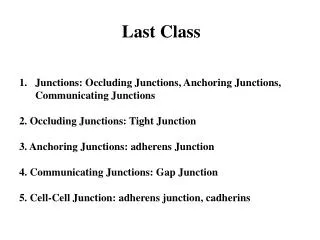
Cell junctions. Intercellular junctions. A. Occluding junctions Tight junctions B. Anchoring junctions Macula adherens (Desmosomes) Zonula adherens Fascia adherens C. Communicating junctions Gap junctions Chemical synapse. Tight junction. Also called as Zona occludens

Cell junctions
E N D
Presentation Transcript
Intercellular junctions A. Occluding junctions • Tight junctions B. Anchoring junctions • Macula adherens (Desmosomes) • Zonula adherens • Fascia adherens C. Communicating junctions • Gap junctions • Chemical synapse
Tight junction • Also called as Zona occludens • Formed by fusion of the plasma membrane • Made up of ridges form each of the cell called cingulin • Located in the apical part of the lateral surface of the cells • Eg: intestinal epithelium, gall bladder, choroid plexus, etc
Functions: • It forms a barrier to the movement of ions and other solute from one side of the cell to the other • It maintains the cell polarity by preventing the movements of the integral proteins
Anchoring junctions Zonula adherens Characterised by local thickening of the adjacent cell membranes of two epithelial cells Functions: Helps the tissue to withstand stress and thus prevents the tearing of the cells
Macula adherens (Desmosomes) • Characterised by a uniform space between opposing cell membranes • Adjacent membranes become thickened and the space in between is filled with filament like material containing Cadherin and some other membrane protein. • Fibrils arise from this portion to the interior of the membrane.
Fascia adherens • They are similar to zonula adherens • Area of attachment are in the form of short strips instead of continuous • Seen in smooth muscle, intercalated discs of cardiac muscle, etc
Gap junction • Characterised by 3-4 channels in between two adjacent cells consisting of assembles of channel protein called connexins • These are present in heart muscles and in smooth muscles
Functions: • Allows rapid transmission of electrical impulse from one cell to another • It permits direct transfer of ions and other molecules like sugar, amino acids between the cells
Cell Communication There are four basic mechanisms for cellular communication: 1. direct contact 2. paracrine signaling 3. endocrine signaling 4. synaptic signaling
Paracrine signaling – signal released from a cell has an effect on neighboring cells
Long-distance signaling Blood vessel Endocrine cell Hormone travels in bloodstream to target cells Target cell (c) Hormonal signaling. Specialized endocrine cells screte hormones into body fluids, often the blood. .
Communication between cells requires: ligand: the signaling molecule receptor protein: the molecule to which the receptor binds -may be on the plasma membrane or within the cell
signal pathways • Signal molecule (ligand) • Receptor • Intracellular signal • Target protein • Response Figure 6-3: Signal pathways
Second Messengers • Many signaling pathways involve small, nonprotein, water-soluble molecules or ions, called second messengers. • These molecules rapidly diffuse throughout the cell. • Second messengers participate in pathways initiated by both G-protein-linked receptors and tyrosine-kinase receptors. • Two of the most important are cyclic AMP and Ca2+.
Hormones act on specific receptors. • The receptors are proteins. • When the Ligand binds the receptor, the receptors sends a signal within the cell to modify the activity of the cell. • Different types of receptors • Intra cellular and cell surface receptors • Ion channel receptors ,enzymatic and G-protein receptors.
GENERAL PRINCIPLES OF CELL COMMUNICATION Extracellular signal molecules bind to specific receptors An intracellular signaling molecule whose concentration increases (or decreases) in response to binding of an extracellular ligand to a cell-surface receptor.
Gate closed Signalmolecule(ligand) Ions Ligand-gated ion channel receptor Plasma Membrane Gate open Cellularresponse Gate close Figure 11.7 • Ion channel receptors