Download
1 / 35
350 likes | 407 Views
Explore the neuromotor events in speech production, focusing on the central and peripheral nervous systems, brain divisions, neural firing, speech-related brain areas, and the crucial link between speech and the peripheral nervous system.

E N D
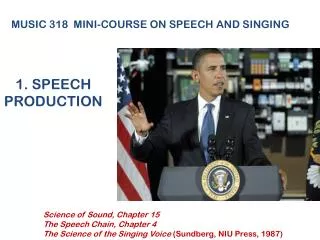
Speech Science PrimerChapter 3: The Raw Materials—Neurology & Respiration
Basic divisions of nervous system • Central nervous system (CNS): Brain and spinal cord • Peripheral nervous system (PNS): All other components, including: • Cranial nerves (exit CNS from brainstem) • Spinal nerves (exit CNS from spinal cord) • Efferent neurons: Nerve impulses from CNS to peripheral parts of the body (motor functions) • Afferent neurons: Nerve impulses from peripheral parts of the body to CNS (sensory functions)
Basic divisions of the brain • Brainstem: Atop spinal cord • Upper brainstem includes thalamus, basal ganglia • Lower brainstem includes pons and medulla oblongata • Cerebellum: Posterior to brainstem • Cerebral hemispheres: Wrap around the brainstem; include areas for higher cognitive function and language
Anatomy & physiology of neurons • Individual neurons contain a cell body plus projections (dendrites, axons) • Axons carry information away from the cell body (efferent) • Dendrites receive information from incoming axons • Firing is “all or nothing”: Stimuli above threshold always generate same response strength • Stronger signals yield more frequent firing (amplitude doesn't change)
Neuronal firing: The action potential • Neuron at rest: • Has negative internal charge • Neuron during firing: • Potassium (K+) exits the neuron • Sodium (Na+) floods into the neuron • Cell interior briefly gains positive charge • Resting negative potential (charge) restored soon after firing
The synapse • At the synapse, the axons of adjacent neurons branch into terminal arbors • The terminal arbors meet the dendrites of the nerve cell receiving incoming stimulation • Neuronal firing releases chemicals (neurotransmitters) into synaptic cleft • Neurotransmitters can either facilitate or inhibit firing in the next neuron(s)
Speech & the CNS • Cortical damage may yield speech or language problems: • Aphasia: Language impairment • Apraxia of speech: Deficits in speech motor programming • Dysarthria: Difficulty with speech movements • Language and speech mainly controlled by left cerebral hemisphere: • Broca's area: Third convolution of left frontal lobe • Wernicke's area: First convolution of left temporal lobe
Motor & sensory areas of the cortex • Motor strip: Frontal lobe • Sensory strip: Parietal lobe • Sensory and motor strips separated by Fissure of Rolando • Representation of the body is upside down in motor and sensory strips • Large amounts of cortex are devoted to the hands and oral (speech) structures
More on laterality • Wada testing: • Used to test for laterality prior to brain surgery • Paralyzes one side of the brain and the side of the body it controls (contralateral) • Language laterality and handedness: • Right-handers: About 96% are left-lateralized for language • Left-handers: About 70% are left-lateralized for language • Some speakers show bilateral organization for language
"You've hissed my mystery lectures" • Spoonerisms: Units of speech/language are exchanged in production (“damp towel” becomes “tamp dowel”) • Errors follow rules: • Consonants only exchange with consonants • Vowels only exchange with vowels • First sounds and syllables are most prone to reversals • Provide evidence that speech is not programmed one word, syllable, or sound at a time
Speech & the peripheral nervous system • Oral and laryngeal structures are innervated mainly by cranial nerves • The respiratory system is innervated by spinal nerves • Efferent impulses interface with muscles in motor units: • An action potential at the motor unit stimulates muscle fibers • Muscle contraction may cause movement of speech structures/articulators or change in muscle tone
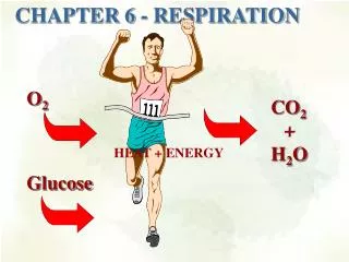
Respiration & speech • All English speech sounds require airflow from the lungs • Airflow forces the vocal folds to vibrate in voiced sounds (phonation) • Obstructing airflow in the upper vocal tract yields supraglottal sound sources (e.g., bursts, frication noise), especially for consonants
Physics of breathing • Expansion of the chest and lungs creates negative pressure (Boyle's law) • Air flows in to equalize the pressure (inhalation) • Contraction of the chest and lungs creates positive pressure • Air flows out (exhalation) • Exhaled airflow is modified for speech production
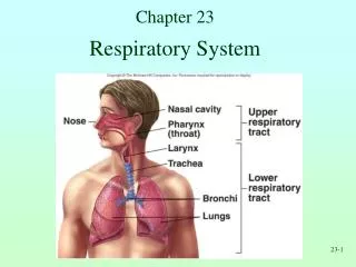
Support structure of respiratory system • Vertebral column • Sternum • Ribs: • Join to vertebral column at back (bony connections) • Upper ribs join sternum at front via cartilage • Lower (floating) ribs connect to vertebrae only
Structure of thoracic cavity • Encircled by bone (ribs, sternum, vertebrae) • Diaphragm forms floor of the thoracic cavity • Pleural linkage connects lungs to rib cage and diaphragm: • Costal (rib) pleura lines rib cage • Pulmonary (visceral) pleura surrounds lungs • Fluid holds the pleural layers together but allows sliding movement • The lungs respond to the expansion and contraction of the rib cage and diaphragm movements
External intercostal muscles • Superficial to internal intercostal muscles • Connect osseous portions of ribs to each other • Run downward toward sternum • Contraction raises and expands rib cage: Inhalation
Internal intercostal muscles • Deep to external intercostal muscles • Run downward away from sternum • Connect both osseous and cartilaginous portions of ribs: • Interosseous portions: Lower and compress rib cage: Exhalation • Interchondral portions: Raise and expand rib cage: Inhalation
Inhalation in quiet breathing • The medulla oblongata sends commands to the respiratory muscles • The diaphragm contracts: • The thoracic cavity expands vertically downward • The external intercostals, interchondral portions of internal intercostals, contract: • The thoracic cavity expands up and out • Lung volume increases because of pleural linkage • Air pressure within the lungs decreases • Air flows in through the nose and mouth
Speech breathing: Overview • More air is typically inhaled than in quiet breathing (especially for loud or long utterances) • Accessory muscles of neck, chest, abdomen, and back may assist in expanding rib cage • Control is more voluntary and conscious than in quiet breathing • Exhalation is slower and takes up more of the respiratory cycle
Passive expiration in quiet breathing • Relaxation of the respiratory muscles with air in the lungs: • Allows the lungs and rib cage to recoil • Respiratory system collapses • Air pressure within the lungs increases • Air flows out • Lungs return to resting volume
Respiratory quantities • Tidal volume: Amount of air exchanged (in and out) during a cycle of quiet breathing • Vital capacity (VC): Amount of air exchanged in maximum inspiration-maximum expiration: • Respiratory volumes often expressed as a percentage of VC (e.g., tidal volume is about 10% of VC) • Resting volume: The respiratory system relaxes at about 40% of VC
Active expiration (speech & singing) • Above resting volume: • Muscles counteract passive collapse of lungs • Inspiratory muscles maintain lungs in expanded state • Slow expiration early during exhalation phase • Below resting volume: • Muscles force respiratory system into compressed state • Expiratory muscles compress thorax and abdomen • Maintain expiration longer
Details of muscle activation for speech • During breathing, both inspiratory and expiratory muscles are active most of the time • The balance between inspiratory and expiratory muscle action changes continuously • The respiratory system maintains fairly constant pressure during speech • Small variations occur to change intensity (e.g., for stressed syllables)
The respiratory system & syllable stress • Increasing subglottal pressure (Ps) yields an increase in intensity (I): • I = Ps3 or Ps4 • Small increases in Ps cause large increases in I • Abdominal and internal intercostal muscles probably raise Ps for stressed syllables • Higher Ps may contribute to other features of syllabic stress: • Higher f0 • Increased duration
Speech breathing & phrasing • Inspirations usually occur at major linguistic boundaries (phrases, sentences) • Long utterances require muscle control to maintain subglottal pressure (Ps) throughout • Utterance requirements affect both inspiratory and expiratory muscle use
Respiratory control in clinical populations • Voice disorders: Improper laryngeal valving may waste exhaled air • Hearing impairment: Poor laryngeal control may again waste air • Motor speech disorders: May affect respiratory muscle coordination