Download
1 / 1
20 likes | 195 Views
INTRAVASCULAR ULTRASOUND EVALUATION OF COMPLEX BIFURCATION LESIONS TREATED WITH TRYTON SIDE-BRANCH STENT IN CONJUNCTION WITH EVEROLIMUS-ELUTING STENTS.

E N D
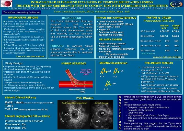
INTRAVASCULAR ULTRASOUND EVALUATION OF COMPLEX BIFURCATION LESIONS TREATED WITH TRYTON SIDE-BRANCH STENT IN CONJUNCTION WITH EVEROLIMUS-ELUTING STENTS D. Trabattoni, MD, FACC; F. Fabbiocchi, MD; P. Montorsi, MD; S. Galli, MD; P. Ravagnani, MD; A. Lualdi, MD; L. Grancini, MD; A.L. Bartorelli, MD, FACC Centro Cardiologico Monzino, IRCCS, University of Milan, Milan - Italy The authors have nothing to disclose BIFURCATION LESIONS TRYTON vs. CRUSH Postprocedural IVUS Data TRYTONstentCHARACTERISTICS BACKGROUND The Tryton Side-Branch Stent was designed to treat complex bifurcation lesions (BL). The results of FIM study demonstrated safety and feasibility and low restenosis rate at 6-month angiographic follow-up. PURPOSE:To evaluate clinical outcome, restenosis rate and ultrasound results after BL treatment with the Tryton Stent. • Cobalt Chromium alloy • Strut thickness= 0.0033” (85 µm) • Stent length= 18 mm • Angles: all • Generous landing zone • (positioning tolerance) • Restenosis of bifurcation lesions remains high even in the DES era, particularly with the two-stent technique • Crush-stent technique offers complete coverage of SB but postprocedural IVUS imaging showed*: • MSA significantly smaller in SB than in MV • SB stent frequently underexpanded, tipically at the ostium • MSA of SB <4 mm2 in 55%, <5 mm2 in 90% • Incomplete SB or MV stent apposition in the crush area (60%) Stent underexpansion not detected by angiography TRYTON N=10 CRUSH* N=15 SB Ostium MSA 4.711.05 4.2±1.0 SB Distal MSA 4.451.1 4.5±2.3 SB stent CSA < 4 mm2 21% 55% SB stent CSA < 5 mm2 66% 90% SB Dmin/D max 0.83 N/A DELIVERYSYSTEM • Rapid exchange catheter • Single-wire tracking • No need for rotational orientation • Low Profile • - 5Fr guide compatible • Balloon Semi-compliant * Non LM lesions *Costa et al. JACC 2005; 46:599-605 Costa et al. JACC 2005; 46:599-605 StudyDesign: MEDINA Classification PRELIMINARY RESULTS HYBRID STRATEGY • Single-center prospective study (n=30 patients) • Six-month angiographic and IVUS F/U • Postintervention and F/U IVUS analysis in both branches • 40-MHz IVUS catheter (BSC) advanced 10 mm beyond • and proximal to the stented segments • IVUS imaging recorded during automated transducer pullback (0.5 mm/s) onto a CD rom for off-line analysis • 11 patients (8 men, 3 women, mean age 64±12 yrs) • 10 LAD-Diag and 1 LCx-OM • All Tryton stents correctly implanted in SB and all DES (Xience V) properly placed in MV • Kissing balloon performed in all cases • 100% angio and procedural success • IVUS imaging of all SB and of 10/11 MV Tryton Side-Branch Stent (BMS) n=9 n=1 1,1,1 1,1,0 n=1 0,1,0 Main-Vessel Stent (DES) • When used in conjunction with a DES, the Tryton stent is associated with good clinical outcome and low restenosis rate • These preliminary IVUS results show: • - Complete coverage of side branch ostium • - Complete stent expansion • - Large final area • High symmetry (Dmin/Dmax) of the Tryton • This may contribute to the low restenosis observed in Tryton I • These preliminary data suggest that the Tryton stent approach provides a reliable and reproducible strategy to stent the SB and its origin C O N C L U S I O N S 6-Month Clinical F/U (n=9) MACE: 1 death (@14 days in recent large anterior STEMI) TLR: 0 TVR: 1 MV (disease progression on LM-LAD) 6-Month angiographic F/U (n= 8 [80%]) In-stent restenosis at 6 months Main Vessel 0% Side branch:0% IVUS IMAGES Post Tryton Stent Implantation 6-mos Follow-up D1 D1 LAD LAD