Download
1 / 1
10 likes | 158 Views
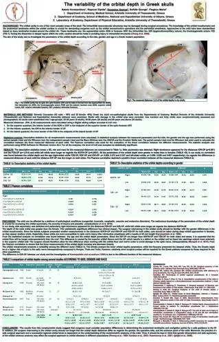
The variability of the orbital depth in Greek skulls Natsis Konstantinos 1 , Repousi Elpida 2 , Noussios George 3 , Sofidis George 1 , Piagkou Maria 2 Department of Anatomy, Medical School, Aristotle University of Thessaloniki, Greece

E N D
The variability of the orbital depth in Greek skulls NatsisKonstantinos1, RepousiElpida2, Noussios George3, Sofidis George1, Piagkou Maria2 • Department of Anatomy, Medical School, Aristotle University of Thessaloniki, Greece • Department of Anatomy, School of Medicine, National and Kapodistrian University of Athens, Greece 3. Laboratory of Anatomy, Department of Physical Education, Aristotle University of Thessaloniki, Greece BACKGROUND: The orbital cavity is one of the most complex areas of the skull. The intraorbital neurovascular structures may be damaged during surgical procedures. The knowledge of the orbital morphometryand the anatomical landmarks within the orbital cavities is crucial for demanding surgeries such as the orbital reconstructive operations and the periorbital anesthesia. Approaches to the orbit have been standardized based on bony landmarks located around the orbital rim. These landmarks are: the supraorbital notch- SON or foramen- SOF, the infraorbital rim- IOR (zygomaticomaxillary suture), the frontozygomatic suture- FZS (FIG.1). During the dissection in deeper layers within the orbit, caution should be made in avoiding injury to intraorbital structures (Cheng et al. 2008). The aim of the study was to investigate the parameters of the orbital depth according to the side, gender, and age in a Greek modern population. Fig 2. The measured distances 1,2,3 of the orbital depths in dry skulls. Fig 1. The orbital cavity has as apex the optic foramen (OF) and its base is formed from the supraorbital rim (SOR), the infraorbital rim (IOR), the frontozygomatic suture (FZS) and the anterior lacrimal crest (SON- superior orbital notch, AEF- anterior ethmoidal foramen, PEF- posterior ethmoidal foramen). MATERIALS AND METHODS: Seventy Caucasian (42 male – 60% and 28 female- 40%) Greek dry adult non-pathological skulls from the Departments of Anatomy, Medical Schools of the Aristotle University (Thessaloniki) and National and Kapodistrian University (Athens) were examined. Skulls with damage in the orbital area were excluded. One hundred and forty orbits were morphometrically assessed and photographed. All skulls were subdivided into 3 age-groups: 20-39 years (9 skulls), 40-59 years (26 skulls) and 60 years and above (35 skulls). Measured distances: (FIG. 2) All measurements were calculated using a Digital sliding calliper accurate to 0.01 mm. • On the superior quadrant, the inferior border of the SON or SOF to the midpoint of the superior border of the optic foramen (OF) • On the inferior quadrant, the IOR to the inferior border of OF • On the lateral quadrant, the inner border of the FZS to the midpoint of the lateral border of OF Statistical analysis:Descriptive statistics for all morphometric measurements were calculated. A statistical analysis between the measured parameters and the side, the gender and the age was performed using the paired sample t-test and the Wilcoxon test, the independent sample t-test and the Mann-Whitney test, the ANOVA and the Kruskal- Wallis test. The paired sample t-test and the Wilcoxon test were used to calculate the correlation between the three measured distances of each orbit. The Pearson correlation was used for the evaluation of the linear correlation between the different measurements. The statistic analysis was performed using SPSS Software for Windows version 20.0. For all the analyses, the level of 0.05 was accepted as statistically significant. SON FZS OF IOR RESULTS: The descriptive statistics (mean ± SD, min and max) for the measured distances are summarized in Table 1a. Side asymmetry was detected. Right dominance appeared for the distances IOR-OF (p=0.001) and the FZS-OF (p= 0.014) and while left orbits were longer as regards the SOF/N-OF (p=0.0001). All the parameters of the orbital depth were greater in males than in females (TABLE 1B). In our study no correlation was found between the orbital depth and the age(right-sided orbits FZS-OF, SOF-OF and IOR-OF- p= 0.628, 0.674 and 0.327 and left-sided orbits, p= 0.296, 0.929 and 0.607 respectively). As regards the differences in measured distances of each orbit the distance IOF-OF was the longer on both sides. The Pearson correlation depicted a positive linear correlation between all the measured distances (TABLE 2). TABLE 1b- Descriptive statistics of the orbital depths according to gender TABLE 1a- Descriptive statistics of the orbital depths TABLE 2 Pearson correlations DISCUSSION: The orbit can be affected by a plethora of pathological conditions (congenital, traumatic, neoplastic, vascular and endocrine disorders). The meticulous knowledge of the parameters of the orbital depth is crucial for accessing the orbital asymmetry, the severity of trauma and possible complications (Ji et al. 2010). In our study, side asymmetry was detected and particularly right dominance appeared for the distances FZS-OF and IOR-OF, while left orbits were longer as regards the distance SOF/N-OF. The depth of the male orbits was greater than the female. This statistically significant difference has clinical impact. The surgeon intervening in the orbital cavity should be familiar with the gender differences in the orbital morphometry. Since the female subjects presented smaller measurements in the distances SOF/N-OF and IOR-OF and FZS-OF for both sides, care should be taken during deep orbital approaches in females, as they appear shallow orbits. Especially, these orbits are more susceptible to optic nerve injury after retrobulbar anesthesia with a needle of 40 mm length (Karampatakis et al. 1998). From the statistical analysis no correlation was found between the age and the orbital depth parameters. Since the IOF-OF are longer than SOF/N-OF and FZS-OF for both sides and sexes, it is expected that the orbital floor will be longer anteroposteriorly than the roof. This difference is caused by the superior orbital fissure and the OF, which results in a defect on the orbital roof as these two openings extend more anteriorly in the superior orbital wall. The surgeon should therefore allow for this difference when working with the orbital floor and roof in order to avoid damage to the optic nerve, intraoperatively (Munguti et al. 2012). From the Pearson correlation is shown that the three measurements of the orbital depth increase and decrease linearly. The orbital morphometry presents a variability among the different races (TABLE 3). The Greeks present the lower orbital depths parameters, while the Kenyans presented the deepest orbits. Thus, the Greeks might be at a higher risk of injury to the optic nerve intraoperatively compared to Kenyan, Thais and Turks. However, relatively short surgical probes will be required for the Greek patients compared to those required for the other populations. The difference in IOR-OF between our study and the investigation of Karampatakis and co-authors (1998) is due to the different borders of the measured distance. TABLE 3 comparison of orbital depths among several studies ACCORDING TO SIDE, GENDER AND RACE REFERENCES Cheng AC, Lucas PW, Yuen HK, Lam DS, So KF. Surgical anatomy of the Chinese orbit. OphthalPlastReconstrSurg 2008;24(2):136-141 Gosavi SN, Jadhav SD, Zambare BR. Orbital morphometry with reference to bony landmarks. Rev Arg de AnatClin 2014; 6(1): 20-25 Hamilton RC. Techniques of orbital regional anaesthesia. Br J Anaesth 1995;75(1):88-92 Ji Y, Qian Z, Dong Y, Zhou H, Fan X. Quantitative morphometry of the orbit in Chinese adults based on a three-dimensional reconstruction method. J Anat. 2010;217(5):501-506 Huanmanop T, Agthong S, Chentanez V. Surgical anatomy of fissures and foramina in the orbits of Thai adults. J Med Assoc Thai 2007; 11: 2383-2391. Hwang K, Baik SH. Surgical Anatomy of the orbit of Korean adults. J CraniofacSurg 1999; 2:129-134 Igbigbi SP, Ebite EL. Orbital Index of Adult Malawians. Internet J. Forensic Med. Toxicol2010; 11: http//www.geradts.com/anil/ij Karakas P, Bozkir MG, Oguz O. 2003. Morphometric measurements from various reference points in the orbit of male Caucasians. SurgRadiolAnat 6: 358-362 Karampatakis V, Natsis K, Gigis P, Stangos NT. Orbital depth measurements of human skulls in relation to retrobulbar anesthesia. Eur J Ophthalmol 1998;8(2):118-120 Karampatakis V, Natsis K, Gigis P, Stangos NT. The risk of optic nerve injury in retrobulbar anesthesia: a comparative study of 35 and 40 mm retrobulbar needles in 12 cadavers. Eur J Ophthalmol 1998;8(3):184-187 Katsev DA, Drews RC, Rose BT. An anatomic study of retrobulbar needle path length. Ophthalmology 1989;96(8):1221-1224 Munguti J, Mandela P, Butt. F. Referencing orbital measures for surgical and cosmetic procedures. Anatomy Journal of Africa. 2012;1(1):40-45 CONCLUSIONS: The results from this morphometric study suggest that surgeons must consider population differences in determining the anatomical landmarks and navigation points for a safe guidance to the OF. In addition, the surgeon intervening in the orbital cavity should not forget that the orbital depth distances differ as regards the gender, the operative side, and the entrance point of the orbit. Moreover, the practice of a safe surgical approach and a successful regional orbital block is dependent on the understanding of the morphometric anatomy of the orbit. Thus, it should be kept in mind that gender dimorphism and side asymmetry of the orbital osseous anatomy may affect the surgical approach to orbital diseases in different populations (Hwang et al., 1999; Karakas et al., 2002; Huanmanop et al., 2007; Igbigbi et al., 2008).