Human Development and Genetics
The development of the embryo-fetus takes place in the uterus and lasts an average of 40 weeks. . Fertilization of the egg by the sperm usually takes place in the fallopian tube.The acrosome of the sperm becomes more fragile (capacitation); the enzymes released will digest the membrane of the egg.

Human Development and Genetics
E N D
Presentation Transcript
1. Chapter 21 Human Development and Genetics
2. The development of the embryo-fetus takes place in the uterus and lasts an average of 40 weeks. Fertilization of the egg by the sperm usually takes place in the fallopian tube.
The acrosome of the sperm becomes more fragile (capacitation); the enzymes released will digest the membrane of the egg.
The diploid number is restored in the zygote.
Both men and women have 22 pairs of autosomes.
Sex chromosomes: XX � XY �
3. Zygote to blastocyst � implantation takes place 5 to 8 days after fertilization Within the fallopian tube the zygote begins a series of mitotic divisions called cleavage: ? two-cell stage ? four-cell stage ? eight-cell stage, and so on.
A morula is a solid sphere of cells.
Blastocyst � the trophoblast (outer cell layer) secretes enzymes to form a crater in the endometrium �
Inner cell mass � embryonic stem cells.
4. Embryo � weeks 1 through 8 of gestation In the embryonic disc, 3 primary germ layers develop (see Table 21�1):
Ectoderm �
Mesoderm �
Endoderm �
By the 8th week (embryo is 1 to 1.5 in. long), all of the organ systems are formed.
5. Embryonic membranes Yolk sac � forms the first blood cells and reproductive stem cells.
Amnion � surrounds the fetus; contains amniotic fluid �
Chorion � develops chorionic villi that will contain blood vessels and will become the fetal portion of the placenta.
6. Fetus � weeks 9 through 40 of gestation The organ systems grow and mature (see Table 21�2).
The growing fetus brings about structural or functional changes in maternal organ systems (see Table 21�3).
Question: Which of the fetal organ systems is the last to become functional?
7. Answer The fetal respiratory system is the last organ system to become functional.
8. Placenta and umbilical cord The placenta is formed by the chorion of the embryo and the endometrium of the uterus.
Fetal blood does not mix with maternal blood in the placenta; fetal capillaries are within maternal blood sinuses. This is the site of exchanges between fetal blood and maternal blood.
The placenta is delivered after the baby and is called the afterbirth.
Question: What are the mechanisms of placental exchange?
9. Answer Mechanisms of exchange in the placenta are diffusion (for exchange of gases and some waste products) and active transport (for exchange of nutrients and some waste products).
10. Placenta and umbilical cord (continued) The umbilical cord connects the fetus to the placenta.
Two umbilical arteries carry blood from the fetus to the placenta.
CO2 and waste products �
O2 and nutrients �
The umbilical vein returns blood from the placenta to the fetus.
11. Placental hormones � the placenta is a temporary endocrine gland Human chorionic gonadotropin (hCG) � secreted by the chorion.
Function: maintains the corpus luteum �
Estrogen and progesterone � secretion begins within 4 to 6 weeks and continues until birth.
Functions of estrogen and progesterone together:
Inhibit secretion of FSH and LH.
Prepare the mammary glands for lactation.
12. Placental hormones (continued) Progesterone inhibits contraction of the myometrium.
Toward the end of gestation, the secretion of progesterone decreases while estrogen secretion remains high �
Relaxin � permits stretching of the pubic symphysis and inhibits contractions of the myometrium.
13. Parturition and labor
Parturition � birth.
Labor � the sequence of events during birth.
First stage � dilation of the cervix �
Second stage � delivery of the infant
Oxytocin �
Third stage � delivery of the placenta.
14. The infant at birth The umbilical cord is clamped and severed.
Increased CO2 stimulates breathing; the lungs are inflated; more blood returns to the left side of the heart.
The foramen ovale closes.
The ductus arteriosus constricts.
The ductus venosus constricts.
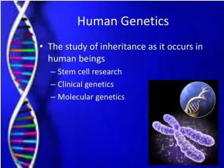
15. Genetics � the study of inheritance Human cells have 46 chromosomes, in 23 homologous pairs.
Homologous pair: a maternal and a paternal chromosome �
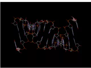
DNA is the hereditary material of chromosomes.
Gene � the genetic code for one protein.
Alleles � the possible ways a gene may be expressed �
16. Genetics (continued) Genotype � the alleles present in the genetic makeup.
Homozygous �
Heterozygous �
Phenotype � the expression (appearance) of the alleles present.
Phenotype is determined by the dominance or recessiveness of the alleles, or the pattern of inheritance involved.
17. Genetics (continued) Dominant-recessive inheritance
A dominant gene will appear in the phenotype of a heterozygous individual.
A recessive gene will appear in the phenotype only if the individual is homozygous.
Example: Rh blood type
Rh (+) is dominant; Rh (�) is recessive.
Questions: What are the possible genotypes for an Rh(+) person? For an Rh(�) person?
18. Answers An Rh (+) person may have a genotype of ++ or +�.
An Rh(�) person will have the genotype � �.
19. Genetics (continued) Multiple allele inheritance
Each gene has more than two possible alleles.
An individual will have only two of these genes.
Example: ABO blood type
An O gene is recessive.
A and B genes are co-dominant.
20. Genetics (continued) Sex-linked inheritance (X-linked)
The genes for these traits are recessive and are found only on the X chromosome; the Y chromosome does not have corresponding genes.
Women with one gene �
Men cannot be carriers �
Example: red-green color blindness
Question: Name two genetic diseases that are inherited this way.
21. Answer Hemophilia and Duchenne�s muscular dystrophy both have a sex-linked pattern of inheritance.
22. Wrap-Up Question Name the part or aspect of development or genetics described.
1. The appearance or expression of a trait
2. Inheritance of hemophilia
3. Having two different alleles for a trait
4. Maternal and paternal chromosome pair
5. Embryo stage implanted in uterus
6. Embryonic part of the placenta
7. Carry blood from fetus to placenta
8. Hormone that facilitates labor
23. Answers 1. The appearance or expression of a trait � phenotype
2. Inheritance of hemophilia � sex-linked
3. Having two different alleles for a trait � heterozygous
4. Maternal and paternal chromosome pair � homologous
5. Embryo stage implanted in uterus � blastocyst
6. Embryonic part of the placenta � chorion
7. Carry blood from fetus to placenta � umbilical arteries
8. Hormone that facilitates labor � oxytocin