Download

1 / 177
1.78k likes | 2.07k Views
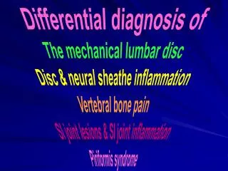
Differential diagnosis of The mechanical lumbar disc Disc & neural sheathe inflammation Vertebral bone pain SI joint lesions & SI joint inflammation Piriformis syndrome. Muscle Testing in Applied Kinesiology.

E N D
Differential diagnosis ofThe mechanical lumbar discDisc & neural sheathe inflammationVertebral bone painSI joint lesions & SI joint inflammationPiriformis syndrome
Muscle Testing in Applied Kinesiology • Research has indicated that the AK muscle test is dependent upon the integrity of the entire neuromuscular complex. • In theory, then, it should be possible with the muscle test to evaluate local and more central control mechanisms.
Muscle Testing in Applied Kinesiology • The dependence of the muscle test on the integrity of the nervous system does not necessarily imply that the act of testing a muscle will immediately evidence abnormalities within that complex.
AK Muscle Testing Designed to Uncover the Cause of the Problem • This is accomplished by a combination of: • Specific muscle testing. • The muscle(s) chosen must be as closely associated with the problem being examined as is possible. • Ideally, the muscles tested will already be weak or over-facilitated in response to the problem. • A specific ‘challenge’ coupled with the muscle test in an attempt to change the muscle response pattern which will evidence the disturbance.
It has been estimated that almost 90% of all individuals will suffer from low back pain at least once in their lives. • 40-50% - Will have pain at least once per year. • 20-30% - Will frequently experience chronic pain. • 2-5% - Will have continual pain.
Wiesel SW (1984) A Study of Computer-Assisted Tomography: The Incidence of Positive CAT Scans in an Asymptomatic Group of Patients, Spine 9(6):549-551. • Often disc herniation is not the origin of pain. • About 40% of adults with an age greater than 40 and the presence of a herniated disc do not have pain.
Kirkaldy-Willis, William MD • The lumbar area consists of three components: • The intervertebral disc • The two zygoapophyseal joints • Any loss of disc height can alter the function of the zygoapophyseal joints. • The posterior joints are extremely rich in nociceptors • Pain can originate from any one or all three of the components
The Evolution of Disc Herniation • Two classic theories for the herniation. • Spontaneous prolaps • With the spine in flexion, the individual attempts to lift a weight which is too heavy and the intradiscal pressure becomes too great and suffers damage. • A gradual prolaps • Repetitive, prolonged stress to the disc fatigues the annulus fibrosis and predisposes it to further damage.
Natural History of the Disc • In most patients, the disc during a period of several months will spontaneously reduce in size. • The reason for this reduction in size is: • A drying out of the discal material. • Why this occurs, at this moment not known.
Theories for the spontaneous reduction of the disc • Reabsorption: • A deficiency in nutrition within the disc caused by a severe loss of bioavailability of nutrients to the disc. • Desiccation (dehydration) – Caused by a deficiency of hydrophilic proteoglycans. • Phagocytosis – Secondary to the inflammatory response during the acute phase of disc herniation.
Low Back Society (2004) • Has concluded that the options available to the patient should include specific manual therapy by a skilled specialist. • Also recommended that manual therapy is made more effectively if combined with other adjunctive methods of treatment: • These would include: TENS, Massage, Ultrasound • Yet is it necessary to consider other alternatives?
Professor Karel Lewitt - Neurologist(Medical University - Prague) • Vertebral “blocks” found together with the herniated disc can stimulate a noted worsening of the condition. • More importantly, research has demonstrated that following a manual therapy to “unblock” the vertebrae involed in the condition, a major portion of patients improve in a significant manner.
Professor Karel Lewitt - Neurologist(Medical University - Prague) • One should almost always attempt a series of manual treatments on patients who suffer from lumbosciatica because it always has value. • But he has also said: This recommendation is dependent upon the application of the correct therapy.
Is There More? • Pain caused by muscle? • Strains (Often a diagnosis used) • Weakness from a deficiency in capacity. • As with ‘Back School’. • Pelvic floor musculature post-partum. • Viscero-somatic reflexes? • For example – the prostate in the male and uterus in the female. • Kidneys – stones and infection. • The intestine – colitis, irritable bowel, dysbiosis. • Postural reflexes caused by mechanical problems far removed from the source of pain. • Cervical lesions
Our Dilemma? • How is it possible in a clinical setting to make an accurate diagnosis between the various elements causing pain?
Our Normal Instruments • Static and motion palpation • Examination specific to manual medicine • Orthopedic exams • Neurologic exams • Ex. - Deep tendon reflexes • Range of motion • Experience • Subjective symptoms of the patient • X-ray and other scanning methods
More Often Than Not – With too little time and too many patients we fall back on habitual methods. • We refer the patient to x-ray and scanning in order to arrive at a diagnosis. • If we don’t have these possibilities? • Deep tendon reflexes (not always significant) • A few orthopedic tests – if time permits • Sensory examination (Can be very subjective) • Manual medicine techniques like motion palpation.
In manual medicine, more often than not, the conclusion is to manipulate the spine How do we confirm our decision to manipulate? • The number of treatments is dependent upon – what?
BJ Palmer, DC • The son of David Daniel Palmer, the man who founded the profession of Chiropractic. “One can have 50 people with back pain walk past you and each one is hit on the backside with a shovel. Twenty-five will probably feel better. This is not an indication of good …manipulative therapy”
How do we measure the effectiveness of our treatment regime? If we are fortunate, the patient may feel a positive change immediately – but this is not always the case. • More often than not we take the ‘wait and see’ attitude. • We fill the patient with platitudes of when the pain will ease and how many treatments they will need, but… • We are too often dependent upon the patient’s response as to pain relief in concluding the validity of the treatment rather than having other, less subjective, methods at hand to use in further evaluation.
How May We Increase Our Diagnostic Ability and Therapeutic Conclusions? • Study neurology. • Learn more orthopedic exam procedures. • Continue to follow every course available in manual medicine. • Work with more experienced colleagues.
Is manipulation alone enough? • Research shows that many patients suffer from musculoskeletal pain due to dysfunction in areas far removed from the source of pain.
Cassidy , Thiel e Kirkaldy-Willis Conclude • Pain from the herniated disc is primarily caused by inflammation. • When other neurologic signs are present they say that these are directly associated with the compressive forces caused by the disc. • Sensory loss (paresthesia). • Motor inhibition. • Loss of muscle strength easily determined using AK methods. • Reduced or loss of deep tendon reflexes.
The basis of all healing arts is to attempt to eliminate the suffering of our patients • Statistically the results do not always speak well for our efforts. • The numbers of the suffering are on a steady increase as is the number of those attempting to offer a therapeutic answer.
George Goodheart DCResults are based on…. • Diagnosis • Diagnosis • Diagnosis
Phase oneStart in the seated position. • It is best to start with testing the gluteus medius as it is innervated by the L5 and S1, primarily, • but also some association with the L4 nerve. • It is important to stabilize the opposite knee to prevent against body rotation of the patient. • In some cases, it will be difficult for the patient to extend the leg as is shown due to pain.
Phase one • The leg may be abducted to a greater degree than is shown. • External rotation of the leg at the foot will help to isolate the gluteus medius from the tensor fascia lata. • The patient may aid against rotation by grasping the table. • Whenever in doubt as to the position of the patient or the test just repeat the testing in a way to eliminate the variable.
Make Note of Any Findings • This is called the ‘neutral position’. • Very often, in cases of discal problems, one of the two glutei will test weak. • Nevertheless, in many patients, the initial testing does not evidence any weakness. This is not a problem.
In the Absence of Obvious Weakness • Continue testing with the patient therapy localizing (TL) to the acupuncture point K-27. • This will evidence any over-facilitation caused by a metabolic condition.
The Home of All Associated Points • Therapy localization to the point K-27 on the same side of the body as the muscle being tested. • This is an important development in AK muscle testing as it enables the examiner to quickly and accurately evaluate for over-facilitated muscle reactions. K-27
Begin Lateral Flexion – Step Two • Lateral flexion is designed to provoke mainly the discal element in the lumbar spine. • It is clear that this movement will also effect the posterior zygoapophyseal (facet) joints, but the test is still primarily for eliciting discal reactions. Lateralflexion
Lateralization • After the first gluteus medius testing in the neutral position it is time to add ‘challenge’ variables. • Lateral flexion as shown is made by asking the patient to lower the shoulder only. • Be on guard not to allow the patient to just lean to the side. This will not put much pressure on the disc. Lateralflexion
Lateralization • Once it has been established that there is no over-facilitation, the testing proceeds without TL to K-27. • If there was weakness in part one, look for strengthening of the muscle. • If no weakness was found in part one, look to create it with the movements. Lateral flexion
Test Both Sides in Lateral Flexion • IMPORTANT • Remember in each of the steps what position of the body either strengthened or weakened the muscle being tested. Medial flexion
Test Both Sides in Lateral Flexion • Testing both sides is important, even if weakness is found upon testing the first gluteus medius muscle. • Bilateral weakness would throw some doubt into using the gluteus medius as an indicator. Medial flexion
Postero-medial/central Lesion • Assume the patient had a weak left gluteus medius in the neutral position. • Should the patient’s muscle strengthen in this position, it indicates that there is a possible postero-medial or central disc lesion right. Medial flexion
Panjabi and WhiteThe Postero-medial/Central Lesion • Panjabi and White used pain as a criteria. • Pain can be a subjective tool and it can be absent even though sciatic involvement is evident. • An increase in pain would be felt if the patient laterally flexes away from the side of sciatica and decrease on ipsilateral flexion. Medial or central lesion
Panjabi and White • The patient must respond with a sense of pain reduction or increase. This brings into the test a great amount of subjectiveness. • The muscle test, even though manual, is much less prone to subjective response than the above.
Postero-medial/central Lesion • A medial/central lesion may also be illustrated here. • If the patient was at first testing strong, but weakens to lateral flexion to the opposite side, the same type of lesion is indicated. Lateral flexion Strong muscle now weakens
Postero-lateral Lesion • A lateral or intraforaminal lesion can be illustrated here. • If the patient was at first testing weak, but strengthens to lateral flexion to the opposite side, the above type of lesion is indicated. Lateral flexion A previously weak muscle strengthens
Panjabi and WhiteThe Lateral or Intraforaminal Lesion • In this instance, the pain is increased on flexion towards the symptomatic side and reduced contralaterally. Lateral lesion
Postero-lateral or Intraforaminal Lesion • Assume the patient had a weak left gluteus medius in the neutral position. • The patient’s muscle will remain weak if not become even weaker when in the presence of a right postero-lateral lesion. Lateral lesion
The Research of Nachemson • Measured the increase in L3/4 lumbar disc in various positions. • This illustrates pressure in the erect position.
The Research of Nachemson • During physical activity. • Note the amount of pressure in the supine, but extension position.
The Research of Nachemson • Note that the seated forward bending and the seated forward bending with weight added create some of the greatest increase in intradiscal pressure. • Therefore, testing the patient while seated provides both a stabile base from which to test and the ‘disc challenge’ we are looking for.
Panjabi and White Central paramedial Lateral
Panjabi and WhiteOnce Again Lateral Paramedial