Download

1 / 51
530 likes | 713 Views
Mammography # 1. Week 2. Mammography Facts. 1 in 8 women who live to 95 will develop breast cancer Most common malignancy in women, only lung cancer kills more women One of the most treatable cancers Before Mammo fewer than 5% of pt’s survived 4 years after diagnosis with a 80% recurrence

E N D
Mammography # 1 Week 2
Mammography Facts • 1 in 8 women who live to 95 will develop breast cancer • Most common malignancy in women, only lung cancer kills more women • One of the most treatable cancers • Before Mammo fewer than 5% of pt’s survived 4 years after diagnosis with a 80% recurrence • With a radical mastectomy survival increased to 40% with a 10% recurrence
Goal of Mammography • Detect cancer before it is palpable • Early detection, diagnosis and treatment is the key to a favorable prognosis
How would your family feel with you missing from the family picture?
How would you feel about your father, brother or mother missing from the family picture?
Anatomy of the Breast • Vary in shape & size • Cone shaped with the post surface (base) overlying the pectoralis & serratus muscles • Axillaries tail extends from lat. base of the breasts to axillaries fossa • Tapers ant. from the base ending in nipple, surrounded by areola
Female Breast • Consists of 15-20 lobes • Divide into several lobules • Lobules contain acini, draining ducts and interlobular connective tissue. • By teenage years each breast contains hundreds of lobules
Lymph Nodes • Lymphatic vessels of the breast drain laterally and medially • Laterally into the axillary lymph nodes (C & D) • 75& drain toward axilla • Medially into the mammary lymph nodes • 25% toward mammary chain (F)
Fibro-glandular Breast • Fibro-glandular • Dense with very little fat • Females 15-30 years of age • Or 30 years or older without children • Pregnant or lactating
Fibro-fatty Breast • Fibro-fatty • Average density • 50% fat & 50% fibro-glandular • Women 30-50 years of age • Or women with 3 or more children
Fatty Breast • Fatty • Minimal density • Women 50 and older (postmenopausal), men and children
Ouch! Why Compression? • Two Reasons: • Decrease thickness of breast tissue • Reduce OID
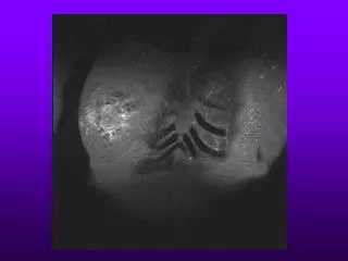
CC positioning • CR Perpendicular • Film tray brought to level of inframammary crease • Wrinkles and folds smoothed out • Compression applied • Markers on axillary side
CC Criteria • No motion • Nipple in profile • All pertinent anatomy demonstrated • Dense areas penetrated • High contrast & optimal resolution • Absence of artifacts • Marker & patient ID visible
MLO positioning • CR & cassette (IR) angled 45 degrees • Top of cassette (IR) at axilla • Compression applied • Nipple in profile • Marker at axilla
MLO criteria • No motion • Pectoral muscle to level of nipple visualized • Breast pulled away from chest wall • Nipple in profile • Dense areas of breast penetrated • High contrast & optimal resolution • Absence of artifacts • Marker & PT ID visible
Complication with Breast Augmentation • Mammography has a 80-90% true positive rate for detecting breast cancer in those women without implants • Decreases to 60% with implants • Because 85% of breast tissue is obscured • More images are needed than the standard two projections • There is a risk of rupturing the implant
Male Mammography • 1300 men get breast cancer per year • 1/3 die • Most are 60 years or older • Nearly all are primary tumors • Symptoms include: • Nipple retraction • Crusting • Discharge • Ulceration
Gynemastia • Benign excessive development of male mammary gland • Occurs in 40% of male cancer pt’s • Survival rates with treatment are 97% for 5 years