Download
1 / 42
490 likes | 809 Views
Molecular basis of dilated cardiomyopathy. Prasad Gunaruwan 21 st June 2011. Introduction. Didactic, regular questions to the audience Pathology of dilated cardiomyopathy Myocyte apoptosis, regulators Structure of the cardiac myocyte Inherited DCM and mechanisms

E N D
Molecular basis of dilated cardiomyopathy Prasad Gunaruwan 21st June 2011
Introduction • Didactic, regular questions to the audience • Pathology of dilated cardiomyopathy • Myocyte apoptosis, regulators • Structure of the cardiac myocyte • Inherited DCM and mechanisms • Extra-cellular components in DCM • Therapeutic potential
Classification of DCM • NEJM pic N Engl J Med 2011;364:1643-56.
Pathology of DCM N Engl J Med 2011;364:1643-56.
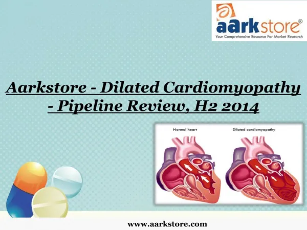
Pathology of DCM • LV dilatation • Systolic dysfunction • Myocyte death and fibrosis • Regardless of etiology this is the end result
Cardiac Myocyte Apoptosis • Myocyte death as a result of necrosis, autophagy (self destruction and recycling of raw material) and apoptosis (programmed cell death) • Apoptosis mediated via external pathway (death ligands binding to cell surface receptors) and internal pathway (via mitochondria and ER) • Internal pathway has many stimuli (hypoxia, DNA damage, radiation, loss of trophic factors, toxins…) • Apoptosis signals kept in check at different levels in the activation cascade
Question to..last row furthest from the door……(ususally Dr Hayes) • What is not a pro-apoptotic intra-cellular component? • Active caspase 3 • Active caspase 9 • Active caspase 12 • Active caspase 8 • X linked Inhibitor of Apoptosis (XIAP)
Evidence for increased apoptosis in DCM • Apoptosis ‘measured’ by caspase activation, mRNA levels of pro-apoptotic components (Bcl-2, Bax, Bcl-xL) and activation of their gene encoding by p-53. • Control apoptosis rate 0.001-0.002% vs 0.08-0.25% in failing hearts. • Loss of myocytes is not debated, but the contribution of apoptosis is controversial. • Causal relationship in mouse models and correlations in human studies
Mouse Model: apoptosis causes DCM • Transgenic mice, cardiac restricted pro-caspase 8 fusion protein expression. • The molecule dimerization and activation could be switched on by a drug. • Within hours of activation death with severe myocyte damage.
These had low levels of caspase and apoptosis (0.023% vs 0.002%). DCM partially arrested with caspase inhibition. J. Clin. Invest. 111:1497–1504 (2003)
Human Studies: apoptosis in DCM (not causal) • 4 clinical groups (normal volume status, AR with preserved EF, AR with reduced EF, severe DCM/AR) • LV biopsies and assay for apoptosis • Patients were on medications
Caspase levels in DCM Am J Cardiol 2009;103:1261–1268)
Ischaemia and apoptosis Am J Physiol Heart Circ Physiol 280: H2313–H2320, 2001.
Other etiology to apoptosis links • Ang-II • Gq transduces Ang-II signals • Gαq (over expression of α subunit) predisposes to transcriptional activation of BH3 like protien Nix/Gp 130/STAT • Eventually Bcl-xL and apoptosis • Severe activation of Nix in pregnant mice: fulminant DCM, inhibitors of Nix prevent the DCM. • Myocardial stretch promotes Titin iso-type switching and p53 activation, which promotes encoding for pro-apoptotic molecules
Question to… Second row furthest from the door.. (usually Prof Fletcher) • Which of the following DCM etiology is yet to have a identified link to apoptosis? • Ischaemic cardiomyopathy • Peripartum cardiomyopathy • Severe aortic regurgitation • Persistantly activated Renin-Ang-Ald system • Alcohol induced DCM
Summary so far… • DCM: LV dilatation, myocyte death/fibrosis, systolic dysfunction. • Multiple etiologies somehow converge on the above final status. • Myocyte apoptosis rates higher in severe DCM. • Some data suggest apoptosis causally linked to DCM. • Some possible pathways to link acquired DCM etiology to apoptosis.
Inherited DCM • Genetic defects resulting in structural defects within the sarcomere • A brief review of normal sarcomeric structure
Lamin A/C DCM • Lamins are ‘structural’ proteins located within the nuclear side of the nuclear membrane • How they cause DCM is still being studied
Lamins: structural vs functional • Important molecules to maintain nuclear envelope structure. • Lamin A has intra-nuclear components that regulates nuclear replication, gene expression and regulates transcription of RNA. • Mutated Lamin A: mis-localises Emerin to ER, and Lamin C to the nucleoplsm. • Mis-localisation of Lamin C into the nucleoplsm seems to be key in DCM. • Mouse models show link to extra-cellular kinase activation
Lamins and Extra-cellular Kinases • Lamin mutation knock-in mice noted to have increased ERK activity prior to DCM • Drugs inhibiting ERK activity delayed DCM onset and severity • ? Lamin mutations may allow RNA transcriptions of these kinases • ? Normal lamins provide substrate for the kinases
Question to.. First row furthest from door.. (usually Dr Collins..) What is incorrect? a) Lamin A/C mutations lead to AutoD and AutoR disorders. b) Lamins are important structural proteins in the sarcolemma. c) Lamin mutation may promote extra-cellular kinase activation. d) Lamins maintain the structure of the nuclear envelope. e) Mis-localisation of Lamin C is associated with DCM.
Summary so far.. • DCM: LV dilatation, systolic dysfunction, myocyte death and fibrosis. • Apoptosis may be a key feature in loss of myocytes. • Links between etiologies and apoptosis. • Any structural protein abnormality in the sarcolemma can lead to DCM. • Lamins are not just structural proteins
Changes in the ECM with DCM • Increased collagen turn-over, ratio of C I:III reduced in DCM • Mast cells and fibroblast activation, increased pro-fibrotic cytokines and growth factors • Loss of collagen cross-linking, mural re-alignment (‘slippage’) • Activated Matrix-Metalloproteinases.
MMP and TIMP • TIMPs are Tissue Inhibitors of MMPs • Ratio between MMP: TIMP in CHF favours persistant activation of MMPs • MMP inhibitors of benefit in mouse models of MI and LVF • Led to the trial of MMP inhibitor in Post STEMI LVF: negative result, blamed on inadequate dosing of the MMP inhibitor.
Summary so far.. • DCM: LV dilatation, myocyte death and fibrosis, systolic dysfunction. • Apoptosis a key in myocyte death, could be causal in DCM • Any structural protein abnormality can lead to DCM • Extra cellular matrix changes also contribute to LV dilatation and systolic dysfunction
What about cardiac stem cells? • Not a lot about what happens to CPC in DCM • Presume they are also affected by the etiological factors and down-stream cascades • Anthracycline induced DCM: Injection of CPC harvested prior to exposure reduce effects of DCM
Therapeutic Potential • Myriad of potential therapeutic targets. • Apoptosis work > 10 yrs, no clinical trials yet. • Some trials on ECM have failed. • ? Kinase inhibitors for lamin DCM