Download
1 / 46
460 likes | 603 Views
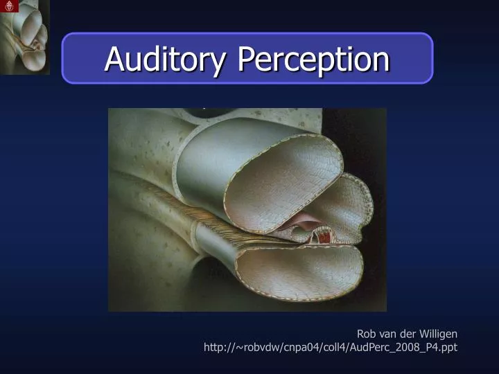
Auditory Perception. Rob van der Willigen http://~robvdw/cnpa04/coll4/AudPerc_2008_P4.ppt. General Outline P4. P4: Auditory Perception. - Cochlear Mechanotransduction. - Neuroanatomical Organization. Sensory Coding and Transduction. Mammalian Auditory Pathway.

E N D
Auditory Perception Rob van derWilligen http://~robvdw/cnpa04/coll4/AudPerc_2008_P4.ppt
General Outline P4 P4: Auditory Perception - Cochlear Mechanotransduction - Neuroanatomical Organization
Sensory Coding and Transduction Mammalian Auditory Pathway Cochlear Mechanotransduction
Sensory Coding and Transduction 6 critical steps
Physical Dimensions of Sound Summary Amplitude - height of a cycle - relates to loudness Wavelength (λ) - distance between peaks Phase (j ) - relative position of the peaks Frequency (f ) - cycles per second - relates to pitch Recapitulation previous lectures
The Adequate Stimulus to Hearing Summary Sound is a longitudinal pressure wave: a disturbance travelling through a medium (air/water) Recapitulation previous lectures http://www.kettering.edu/~drussell/demos.html
The Adequate Stimulus to Hearing Summary Compression Duration Decompression Particles do NOT travel, only the disturbance Particles oscillate back and forth about their equilibrium positions Compression Distance from source Recapitulation previous lectures http://www.glenbrook.k12.il.us/GBSSCI/PHYS/Class/sound/u11l2a.html
Sensory Coding and Transduction A Sensor Called Ear
Sensory Coding and Transduction Peripheral Auditory System Outer Ear: - Extents up to Eardrum - Visible part is called Pinna or Auricle - Movable in non-human primates - Sound Collection - Sound Transformation Gives clues for sound localization
Sensory Coding and Transduction Peripheral Auditory System +60 +40 +20 Elevation (deg) 0 -20 -40 Frequency The Pinna creates Sound source position dependent spectral clues. “EAR PRINT”
Amplitude (dB) Sensory Coding and Transduction Peripheral Auditory System In humans mid-frequencies also exhibit a prominent notch that varies in frequency with changes in sound source elevation (6 – 11 kHz) Elevation +60 +40 +20 Elevation (deg) 0 -20 -40 Frequency kHz
Sensory Coding and Transduction Peripheral Auditory System Barn Owls have Asymmetric Ears and Silent Flight. One ear points upwards, the other downwards.
Sensory Coding and Transduction Peripheral Auditory System Middle Ear: (Conductive hearing loss) - Mechanical transduction (Acoustic Coupling) - Perfect design for impedance matching Fluid in inner ear is much harder to vibrate than air - Stapedius muscle: damps loud sounds Three bones (Ossicles) A small pressure on a large area (ear drum) produces a large pressure on a small area (oval window)
Sensory Coding and Transduction Peripheral Auditory System Inner Ear: The Cochlea is the auditory portion of the ear Cochlea is derived from the Greek word kokhlias "snail or screw" in reference to its spiraled shape, 2 ¾ turns, ~ 3.2 cm length (Humans)
Sensory Coding and Transduction Peripheral Auditory System The cochlea’s core component is the Organ of Corti, the sensory organ of hearing Cochlear deficits cause Sensorineural hearing loss Its receptors (the hair cells) provide the sense of hearing
Sensory Coding and Transduction Peripheral Auditory System The Organ of Corti mediates mechanotransduction: The cochlea is filled with a watery liquid, which moves in response to the vibrations coming from the middle ear via the oval window. As the fluid moves, thousands of hair cells are set in motion, and convert that motion to electrical signals that are communicated via neurotransmitters to many thousands of nerve cells.
Sensory Coding of Sound Cochlear anatomy
Sensory Coding of Sound Cochlear anatomy (straightened)
Sensory Coding of Sound Tonotopic coding Pressure waves distort basilar membrane on the way to the round window of tympanic duct: - Location of maximum distortion varies with frequency of sound - Frequency information translates into information about position along basilar membrane
Sensory Coding of Sound Travelling Wave Theory Periodic stimulation of the Basilar membrane matches frequency of sound Travelling wave theory von Bekesy: Waves move down basilar membrane stimulation increases, peaks, and quickly tapers Location of peak depends on frequency of the sound, lower frequencies being further away
Sensory Coding of Sound Cochlear Fourier Analysis High f Periodic stimulation of the Basilar membrane matches frequency of sound Med f Location of the peak depends on frequency of the sound, lower frequencies being further away Low f BASE APEX Position along the basilar membrane
Sensory Coding of Sound Place Theory Travelling wave theory von Bekesy: Waves move down basilar membrane Location of the peak depends on frequency of the sound, lower frequencies being further away Location of the peak is determined by the stiffness of the membrane
Sensory Coding of Sound Sensory Input is Tonotopic Thick & taut near base Thin & floppy at apex TONOTOPIC PLACE MAP LOGARITHMIC: 20 Hz -> 200 Hz 2kH -> 20 kHz each occupies 1/3 of the basilar membrane
Sensory Coding of Sound Sensory input is tonotopic
Sensory Coding of Sound Sensory Input is Non-linear The COCHLEA: Decomposes sounds into its frequency components Represents sound TONOTOPICALLY Has direct relation to the sounds spectral content Has NO linear relationship to sound pressure Has NO direct relationship to the sound’s location in the outside world
Cochlear nonlinearity Active processing of sound CF= 9 kHz OUTPUT Response in dB ~4.5kHz INPUT level (dB SPL) BM input-output function for a tone at CF (~9 kHz, solid line) and a tone one octave below (~4.5 kHz) taken from the iso-intensity contour plot.
Cochlear nonlinearity Active processing of sound Effects of an “active”cochlea Iso-level curves show sharp tuning at low sound levels, broader tuning at high levels. Response is strongly compressive around the so-called characteristic frequency (CF). Requires functioning outer hair cells. The response of the BM at location most sensitive for ~ 9 KHz tone (CF). The level of the tone varied from 3 to 80 dB SPL (iso-intensity contours).
Cochlear nonlinearity Active processing of sound CF= 9 kHz OUTPUT Response in dB ~4.5kHz INPUT level (dB SPL) Frequency [kHz] The response of the BM at location most sensitive for ~ 9 KHz tone (CF). The level of the tone varied from 3 to 80 dB SPL (iso-intensity contours). BM input-output function for a tone at CF (~9 kHz, solid line) and a tone one octave below (~4.5 kHz) taken from the iso-intensity contour plot.
Cochlear nonlinearity No nonlinearity post mortem Rugero et al. (1997) 1) Reduced gain: Higher thresholds in quiet; loss of audibility as measured with pure-tone audiogram 2) Loss of nonlinearity: Reduced dynamic range; quiet sounds lost but loud sounds just as loud: Loudness Recruitment Basilar-membrane intensity-velocity coding functions for a chinchilla using a tone at the 10 kHz GAIN equals DAmplitude of motion divided by D Amplitude of stimulus pressure
The Auditory Nerve FTC versus FRC FTC data indicate the characteristics of the cochlea from which has been eliminated the non-linear response characteristics of the cochlear nerve excitation process. Response Rate versus Frequency Curve (FRC) FRC data indicate the limits which may be set upon the central representation of the cochlear filtering by the non-linear rate behavior of the cochlear fibers.
The Auditory Nerve Frequency Selectivity: CF & place theory Place Theory: Place of maximum vibration along basilar membrane correlates with the place of the Tuning curve (or FTC=Frequency Threshold Curve) along the frequency axis. Shown are tuning curves measured by finding the pure tone amplitude that produces a criterion response in an 8th nerve fiber (cat). Tuning curves for four different fibers (A-D) are shown.
Cochlear nonlinearity OHC motor driven by the Tectorial membrane A virtuous loop.Sound evoked perturbation of the organ of Corti elicits a motile response from outer hair cells, which feeds back onto the organ of Corti amplifying the basilar membrane motion.
The Problem of Hearing “Sound has no dimensions of space, distance, shape, or size; and the auditory periphery of all known vertebrates contains peripheral receptors that code for the parameters of the sound pressure wave rather than information about sound sources per se.” William A. Yost (Perceiving sounds in the real world, 2007; p. 3461)
The Problem of Hearing Tonotopie blijft in het auditief systeem tot en met de auditieve hersenschors behouden. “De samenstelling van een geluid uit afzonderlijke tonen is te vergelijken met de manier waarop wit licht in afzonderlijke kleuren uiteenvalt wanneer het door een prisma gaat .” John A.J. van Opstal (Al kijkend hoort men, 2006; p. 8)
The Problem of Hearing Mapping can be an important clue to the function of an area. If neurons are arrayed according to the value of a particular parameter, then that property might be critical in the processing performed by that area. Neurons within a brain area may be organized topographically (or in a map), meaning that neurons that are next to each other represent stimuli with similar properties. Neurons do not need to be arranged topographically along the dimensions of the reference frame that they map, even if its neurons do not form a map of that space.
The Problem of Hearing Problem I: Sound localization can only result from the neural processing of acoustic cues in the tonotopic input! Problem II: How does the auditory system parse the superposition of distinct sounds into the original acoustic input?
Sensory Coding of Sound Outer Hair cells Organ of Corti Inner Hair cell Auditory nerve Basilar Membrane Summary
NEXT WEEK Cochlear Innervation & Auditory Nerve Mechanotransduction: Step 5: Vibration of basilar membrane causes vibration of hair cells against Tectorial membrane (TM): • Movement displaces stereocilia/kinocilia, • opens ion channels in hair cell membranes Rush of ions depolarizes hair cells, which initiates the release of neurotransmitters
NEXT WEEK Cochlear Innervation & Auditory Nerve Neural responses in the AN: Step 6 Information about region and intensity of cochlear stimulation is relayed to CNS over cochlear branch of vestibulocochlear nerve (VIII): Called the auditory nerve (AN): Has sensory neurons in spiral ganglion of cochlea Carries neural information to cochlear nuclei (CN) of midbrain for distribution to other (more higher) brain centers.
NEXT WEEK type 1 type 2 Cochlear Innervation & Auditory Nerve Outer hair cells: Primarily receiving efferent inputs. Inner hair cells: Main source of afferent signal in auditory nerve. (~ 10 afferents per hair cell) Type I neurons (95% of all ganglion cells) have a single ending radially connected to IHCs. Type II small, unmyelinated neurons spiral basally after entering the organ of Corti and branch to connect about ten OHCs, in the same row.