Download
1 / 58
590 likes | 713 Views
Understand the Gell-Coombs classification system for hypersensitivity reactions, including Type I (Anaphylaxis), Type II (Cytotoxic), Type III (Immune Complex), and Type IV (Cell-Mediated). Explore allergic reactions, mechanisms, testing, desensitization, and treatment options.

E N D
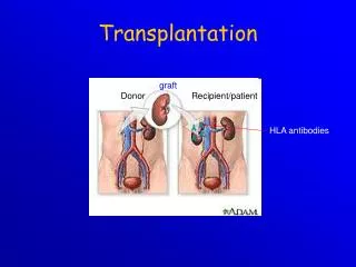
Transplantation M. S. Tvorko
Immunopathology Hypersensitivity Autoimmunity Immunodeficiency
Peter Gell and Robert Coombs developed a classification system for reactions responsible for hypersensitivities in 1963. Their system correlates clinical symptoms with information about immunologic events that occur during hypersensitivity reaction. The Gell-Coombs classification system divides hypersensitivity into four types: Type I (Anaphylaxis) Hypersensitivity Type II (Cytotoxic) Hypersensitivity Type III (Immune Complex) Hypersensitivity Type IV (Cell-Mediated) Hypersensitivity Allergic reactions are subdivided intotwo groups: (1) immediateand (2) delayed reactions,although it is difficult to draw a strict distinction between them. Allergic reactions of immediate action are associated with B-lymphocytes and antibodies circulating in the blood, allergic reactions of delayed action with T-lymphocytes.
TYPE I: IMMEDIATE (ANAPHYLACTIC) HYPERSENSITIVITY Allergen presented to TH2 cells which provide cytokine signals to B cells to produce IgE Ig E binds to mast cells Cross linking of IgE by subsequent exposure to allergen causes mast cell degranulation Mast cells display a high affinity receptor for IgE IgE is synthesised in response to certain antigens (allergens) Allergens are deposited on mucous membranes and taken up and processed by antigen presenting cells (e.g. Dendritic cells or B cells) Mast cell degranulation is the major initiation of the acute allergic reaction Mast cell mediators include histamine, heparin and other factors These cause, mucus secretion, vasodilation and oedema
Mast cell mediators include pre-formed and newly formed mediators • Pre-formed mediators include : histamine, heparin and neutral protease • Newly formed mediators include leukotrienes, prostaglandin D2 and platelet activating factor
Skin testing can be useed for identify the allergen responsible for allergies. These tests involve inoculating small amounts of suspect allergen into the skin. Sensitivity to the allergen is shown by a rapid inflammatory reaction characterizide by redness, swelling, and itching at the site of inoculation
Desensitization.Major manifestations of anaphylaxis occur when large amounts of mediators are suddenly released as a result of a massive dose of antigen abruptly combining with IgE on many mast cells. This is systemic anaphylaxis, which is potentially fatal. Desensitization can prevent systemic anaphylaxis. Acute desensitizationinvolves the administration of very small amounts of antigen at 15-minute intervals. Antigen-IgE complexes form on a small scale, and not enough mediator is released to produce a major reaction. This permits the administration of a drug or foreign protein to a hypersensitive person, but hypersensitivity is restored days or weeks later. Chronic desensitizationinvolves the long-term weekly administration of the antigen to which the person is hypersensitive. This stimulates the production of IgG-blocking antibodies in the serum, which can prevent subsequent antigen from reaching IgE on mast cells, thus preventing a reaction.
Type II Mechanism • Antibodies bind to cell surface • Phagocytes bind to the antibody via their Fc receptor • Phagocytosis of target cell • Antibody binding also activates complement via the classical pathway • Complement mediated cell lysis • Antibody mediated hypersensitivity • Antibody directed against membrane and cell surface antigens (autoantibodies) • Antigen-antibody reactions activate complement producing membrane damage • Examples include: transfusion reactions and haemolytic disease of the newborn
Type III Hypersensitivity Normally immune complexes are degraded by phagocytosis, particularly in the liver and spleen Excessive immune complex formation results in deposition in the tissues, particularly arterioles, kidney and joints Immune complex mediated Excessive formation of immune complexes e.g. persistent low-grade infection, repeated inhalation of antigens Examples of Type III hypersensitivity include: Farmers lung, immune complex glomerulonephritis Complexes induce platelet aggregation and complement activation Attempted phagocytosis causes enzyme release and results in tissue damage
RF is capable of self-associating into immune complexes These immune complexes may fix complement and activate additional inflammatory processes Small immune complexes may directly activate macrophages to produce proinflammatory cytokines by binding to macrophage-surface receptors
Type IV Hypersensitivity • Delayed type hypersensitivity • Takes more than 12 hrs to develop after antigenic challenge • Examples include: contact dermatitis and tuberculin reaction • Antigens include large molecules or small molecules (haptens) linked to carrier molecules
APC resident in the skin process antigen and migrate to regional lymph nodes where they activate T cells • Sensitised T cells migrate back to the the skin where they produce cytokines which attract macrophages which cause tissue damage
Autoimmunity • Autoimmunity is a reaction of the immune system to the bodies own tissues • Self molecules are recognised as antigens due to a breakdown of self-tolerance • Antibodies (autoantibodies) react against these components • Includes organ-specific and non-organ specific diseases
CLASSIFICATION OF AUTOIMMUNE DISEASES • Organ Specific • Insulin dependent diabetes mellitus (IDDM) / Type I) • Grave’s disease • Goodpasture’s syndrome • Myasthenia gravis • Systemic • Systemic lupus erythematosus • Rheumatoid arthritis • Multiple sclerosis • Sjogren’s syndrome
Autoantibodies • ANA (antinuclear antibodies): SLE Anti-ds DNA: SLE • Anti-histone: drug-induced SLE • Anti-IgM (rheumatoid factor): part of RA • Anti-neutrophil: vasculitis • Anti-centromere: CREST - scleroderma • Anti-mitochondrial: primary biliary cirrhosis • Anti-basement membrane: Goodpasture’s (renal, lung) • Anti-epithelial cell: pemphigus vulgaris • Anti-gliadin (not an autoantibody): celiac disease, dermatitis herpetiformis
Systemic Lupus Erythematosus • Chronic, systemic inflammatory disease caused by immune complex formation. • The word "systemic" means the disease can affect many parts of the body. • Pathophysiology associated with clinical features secondary to immune complexes depositing in tissues resulting in inflammation. • Parts of the body affected include: the joints, skin, kidneys, heart, lungs, blood vessels, and brain.
SLE Butterfly Rash • The source of the name "lupus" is unclear. All explanations originate with the characteristic butterfly-shaped malar rash that the disease classically exhibits across the nose and cheeks. • Stranger still, is the account that the term "Lupus" didn't come from latin at all, but from the term for a French style of mask which women reportedly wore to conceal the rash on their faces
Effector mechanisms • Autoantibodies to many autoantigens • Most common autoantibody is to ds-DNA • Immune complex deposition on basement membranes with complement activation and inflammation • Laboratory diagnosis • Anti-nuclear antibody (ANA) • HEp-2 cells • Homogeneous pattern and titer > 1:160 • Anti ds-DNA • Crithidia lucilliae • C3 level
RHEUMATOID ARTHRITIS (RA) • Characterized by inflammation of synovial membrane of joints and articular surfaces of cartilage and bone • Vasculitis is a systemic complication • Affects 3% to 5% of U.S. population • Female to male ratio of 3:1 • HLA DR4 is genetic risk factor
Effector mechanism • CD4 T cells, activated B cells, macrophages and plasma cells • 85% of patients have rheumatoid factor • Rheumatoid factor • IgM, IgG and IgA specific for IgG • Immune complex formation exacerbates inflammation • Laboratory diagnosis • Rheumatoid factor • Anti-cyclic citrulinated peptide • C-reactive protein (CRP)