Download
1 / 60
730 likes | 1.25k Views
GENERAL ASPECTS OF BACTERIAL PATHOGENESIS . Faculty: Dr. Alvin Fox. Suggested reading: Murray, Third edition Chapters 9 & 19. Key Words. Pathogen/Epidemic Adhesion Koch’s postulates Penetration Normal flora Invasiveness/spread

E N D
GENERAL ASPECTS OF BACTERIAL PATHOGENESIS Faculty: Dr. Alvin Fox Suggested reading: Murray, Third edition Chapters 9 & 19
Key Words Pathogen/Epidemic Adhesion Koch’s postulates Penetration Normal flora Invasiveness/spread Infection Extra/intra cellular pathogen Infectious diseases Capsule Compromised host Exotoxin Opportunistic infection Endotoxin Nosocomial Immunopathology Transmission Autoimmunity
Overview Pathogenesis is a multi-factorial process which depends on the immune status of the host, the nature of the species or strain (virulence factors) and the number of organisms in the initial exposure.
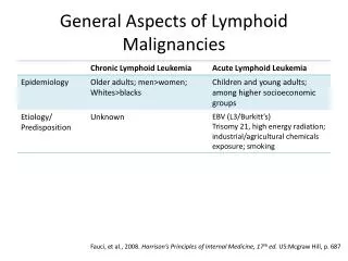
Pathogen/epidemic Limited number of bacterial species responsible for most infectious diseases in healthy individuals. Due to the success of vaccination, antibiotics, and effective public health measures epidemics were felt to be a thing of the past. The development of antibiotic resistant organisms, has changed this situation.
Koch's postulates (modified) 1. The organism must always be found in humans with the infectious disease but not found in healthy ones. 2. The organism must be isolated from humans with the infectious disease and grown in pure culture. 3. The organism isolated in pure culture must initiate disease when re-inoculated into susceptible animals. 4. The organism should be re-isolated from the experimentally infected animals.
Postulates 3. and 4. are important in definite proof of the role of agent in human disease. However, this depends on the ability to develop animal models that resemble the human disease. In many cases such models do not exist.
Opportunistic infections • All humans are infected with bacteria (the normal flora) living on their external surfaces (including the skin, gut and lungs).
Opportunistic infections • Common bacteria found in the normal flora include: • Staphylococcus aureus, S. epidermidisPropionibacterium acnes (skin). • Bacteroides and Enterobacteriaceae, found in the intestine (the latter in much smaller numbers).
Opportunists from the environment • We are constantly also exposed to bacteria including: • air, • water • soil • food.
Normally due to our host defenses most of these bacteria are harmless. In compromised patients, whose defenses are weakened, these bacteria often cause opportunistic infectiousdiseases when entering the bloodstream (after surgery, catheterization or other treatment modalities). When initiated in the hospital, these infectious diseases are referred to as nosocomial.
Transmission Specific bacterial species (or strains within a species) initiate infection after being transmitted by different routes to specific sites in the human body. For example, bacteria are transmitted in airborne droplets to the respiratory tract, by ingestion of food or water or by sexual contact.
Adhesion Bacterial infections are usually initiated by adherence of the microbe to a specific epithelial surface of the host. Otherwise the organism is removed e.g. by peristalsis and defecation (from the gut) ciliary action, coughing and sneezing (from the respiratory tract) or urination (from the urogenital tract). Adhesion is not non-specific "stickiness". Specific interactions between external constituents on the bacterial cell (adhesins) and on the host cell (receptors) occur i.e. an adhesin-receptor interaction.
Example of adhesin S. pyogenes has surface fimbriae which contain two major components the M protein and lipoteichoic acid. The protein fibronectin binds to epithelial cells and fatty acid moieties of lipoteichoic acid in turn interact with fibronectin.
Distinct adhesins can be found within the same organsim. Among the most thoroughly studied pili are those of uropathogenic E. coli. Certain adhesins present on the tips of fimbriae of E. coli facilitate binding to epithelial cells. Type 1 fimbriae bind to mannose containing receptors. While P fimbriae allow binding to galactose containing glycolipids (e.g. cerebrosides) and glycoproteins present on epithelial cells. They are referred to as "P" fimbriae since they were originally shown to bind to P blood group antigens on human erythrocytes.
Penetration and spread Some bacterial pathogens reside on epithelial surfaces e.g. Vibrio cholerae. Other species are able to penetrate these barriers but remain locally. Others pass into the bloodstream or from there onto other systemic sites. This often occurs in the intestine, urinary tract and respiratory tract, and much less commonly through the skin.
For example, Shigella penetrates by activating epithelial cells of the intestine to become endocytic; the Shigella do not usually spread into the bloodstream. In other cases, bacteria (e.g. Salmonella typhi) pass through epithelial cells into the bloodstream. Thus, invasion can refer to the ability of an organism to enter a cell, although in some instances it can mean further passage into the systemic vasculature.
Borrelia burgdorferi is transmitted into the bloodstream through the skin by a tick bite.
Certain degradative exotoxins secreted by some bacteria (e.g. hyaluronidase or collagenase) can loosen the connective tissue matrix increasing the ease of passage of bacteria through these sites.
Survival in the host Many bacterial pathogens are able toresist the cytotoxic actionof plasma and other body fluids involving antibody and complement (classical pathway) or complement alone (alternate pathway) or lysozyme. Killing of extracellular pathogens largely occurs within phagocytes after opsonization (by antibody and/or complement) and phagocytosis. Circumvention of phagocytosis by extracellular pathogens is thus A major survival mechanism. Capsules (many pathogens), protein A (S. aureus) and M protein (S. pyogenes) function in this regard.
Protein A • A surface constituent and a secreted product of • S. aureus. • Binds to the Fc portion of immunoglobulins. • Activates the classical complement cascade. • Phagocytosis occurs after binding of the • opsonized bacteria to receptors for the Fc • portion of IgG or C3 regions.
Protein A is anti-complementary (the complement cascade is activated, depleting complement levels). Protein Ainhibits the interaction of bacteria (via bound complement) with C3 receptors. Free protein A binds to the Fc portion of IgG, preventing phagocytosis via Fc receptors due to steric hindrance.
Peptidoglycan • Can activate the alternate complement cascade. • In S. pyogenes, peptidoglycan is sufficiently • exposed that it is able to bind complement.
M protein The M protein of group A streptococci is the anti-phagocytic component of the fimbriae. M protein binds fibrinogen from plasma which blocks complement binding to the peptidoglycan preventing phagocytosis.
Intracellular pathogens (both obligate and facultative) must be able to avoid being killed within phagolysozomes. This can occur from by-passing or lysing these vesicles and then residing free in the cytoplasm. Alternatively, they can survive in phagosomes (fusion of phagosomes with lysosomes may be inhibited) or the organism may be resistant to degradative enzymes (if fusion with lysosomes occurs).
Tissue Injury • Bacteria cause tissue injury primarily by several • distinct mechanisms involving: • exotoxins • endotoxins and non-specific immunity • (“no antigen”) • “antigen” specific humoral and cell mediated • immunity
Exotoxins Many bacteria produce proteins (exotoxins) that modify, by enzymatic action, or by other means destroy cellular structures. Effects of exotoxins are usually seen acutely, since they are sufficiently potent that serious effects (e.g. death) often result.
Examples of exotoxin produced diseases are botulism, anthrax, cholera and diphtheria. If the host survives the acute infection, neutralizing antibodies (anti-toxins) are often elicited that neutralize the affect of the exotoxin.
(1)Exotoxins which act onextracellular matrix of connective tissue. • e.g. Clostridium perfringenscollagenase • Staphylococcus aureushyaluronidase
Damage to the connective tissue matrix (by hyaluronidase and collagenase) can "loosen up" the tissue fibers allowing the organism to spread through the tissues more readily. Also included in this group is the exfoliatin of Staphylococcus aureus which causes separation of the layers within the epidermis and is the causative agent of scalded skin syndrome in the newborn.
(2)Exotoxins which have a cell binding "B" component and an active "A" enzymatic component (A-B type toxins) • A - B Toxins: • Consist of two components • One binds to cell surfaces • One passes into the cell membrane or cytoplasm where • it acts • cholera and diphtheria toxins.
A-B toxins include: a) Those with ADP-ribosylating activity 1) cholera toxin, 2) E. coli heat labile toxin 3) Pseudomonas aeruginosa toxin 4) diphtheria toxin b) Those with a lytic activity on 28S rRNA 1) shiga 2) shiga-like (vero) toxins c) Those with a partially characterized site of action 1) botulinum toxin 2) tetanus toxin 3) anthrax lethal toxin
A - B Toxins: (i) ADP-ribosylating exotoxins
Diphtheria toxin (produced by Corynebacterium diphtheriae) is coded by the phage tox gene. The toxin is synthesized as one polypeptide chain and readily nicked into two chains held together by a disulfide bond. B binds to cells and A has the enzymatic activity. A is endocytosed and from the endosome passes into the cytosol. Diphtheria toxin ADP-ribosylates elongation factor (EF2) in ribosomes, thus inhibiting protein synthesis. Pseudomonas exotoxin A has an similar mode of action to diphtheria toxin.
Cholera toxin has several subunits which form a ring with one A subunit inserted in the center. 1) B binds to gangliosides on the cell surface and appear to provide a channel through which A penetrates. 2) A1 is formed by proteolytic cleavage and after internalization ADP-ribosylates a cell membrane regulator complex (using NADH as a substrate), in turn causing activation of adenylate cyclase. Activation of adenylate cyclase causes an increase in cyclic AMP production with resulting decrease in sodium chloride uptake from the lumen of the gut and active ion and water secretion with a watery diarrhea resulting. E. coli labile toxin has a similar mode of action.
Shiga toxins (chromosomally encoded) are involved in the pathogenesis of shigellosis. Shiga-like toxins (phage encoded) are produced by enterohemorraghic E. coli. They share a common mode of action. A fragment of the A subunit passes to the ribosome where it has N-glycosidase activity on a single adenosine residue; i.e. the bond between the base and ribose is lysed. Diarrhea results not from active ion/water secretion, but poor water absorption due to death of epithelial cells from inhibition of protein synthesis.
Botulinum neurotoxins, tetanospasmin and the lethal toxin of B. anthracis appear to be A-B type exotoxins.
Botulinum toxin acts by causing inhibition of release of acetylcholine at the neuromuscular junction. Tetanus toxin is taken up at neuromuscular junctions and transported in axons to synapses. It then acts by inactivating inhibitory neurons.
The exotoxins of tetanus and botulism appear to have B components, but the mode of action of their A subunits are not known. The B component of lethal toxin of B. anthracis is the protective antigen; interestingly, this also serves as the B subunit for edema toxin.
(3) Membrane damaging toxins • Proteases • Phospholipases • Detergent -like action
Membrane Damaging Toxins: These toxins enzymatically digest the phospholipid (or protein) components of membranes or behave as detergents. In each case holes are punched in the cell membrane and the cytoplasmic contents can leach out.
The phospholipase ("toxin") of C. perfringens is an example of a membrane damaging toxin. It destroys blood vessels stopping the influx of inflammatory cells. This also helps create an anaerobic environment which is important in the growth of this strict anaerobe. The delta toxin of S. aureus is an extremely hydrophobic protein that inserts into cell membranes and is believed to have a detergent-like action.
Despite the advances of the antibiotic era, around 200,000 patients will develop Gram negative sepsis each year of whom around 25-40% will ultimately die of septic shock. Septic shock involves hypotension (due to tissue pooling of fluids), disseminated intravascular coagulation and fever which is often fatal from massive system failure. Septic shock results in a lack of effective oxygenation of sensitive tissues such as the brain. There is no effective therapy to reverse the toxic activity of lipid A or peptidoglycan in patients.
Endotoxins are toxic components of the bacterial cell envelope.The classical and most potent endotoxin is lipopolysaccharide. However, peptidoglycan displays many endotoxin-like properties.
Certain peptidoglycans are poorly biodegradable and can cause chronic as well as acute tissue injury. Endotoxins are "non-specific" inciters of inflammation. For example, cells of the immune system and elsewhere are stimulated to release cytokines (including interleukin 1 and tumor necrosis factor).
Endotoxins also activate the alternate complement pathway. The production of cytokines results in attraction of polymorphonuclear cells into affected tissues. PG and LPS and certain other cell wall components (e.g. pneumococcal teichoic acid) are also activators of the alternate complement cascade.
Many bacteria will bind complement, encouraging their uptake and killing by phagocytes in the absence of antibody. Certain complement by-products are also chemoattractants for neutrophils.