Download
1 / 54
600 likes | 884 Views
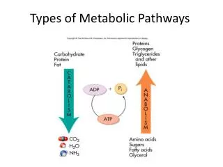
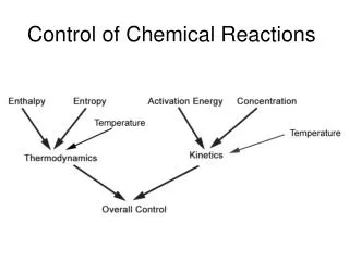
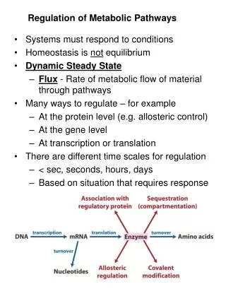
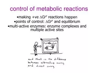
control of metabolic reactions. making +ve D G o ‘ reactions happen points of control: D G o ‘ and equilibrium multi-active enzymes: enzyme complexes and multiple active sites. reactions with +ve D G o ‘ can occur by:. coupling with a reaction with –ve D G o ‘

E N D
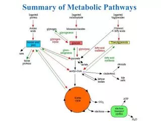
control of metabolic reactions making +veDGo‘reactions happen points of control: DGo‘ and equilibrium multi-active enzymes: enzyme complexes and multiple active sites
reactions with +ve DGo‘ can occur by: • coupling with a reaction with –ve DGo‘ • –ve physiological DG due to cellular low ratio [products]/[reactants]
Thus, ATP ADP +Pi(DG<0) is coupled with non-spontaneous reactions (DG>0) Glucose glucose-6-P + H20 DG = 13.8 kJ.mol-1 hexokinase DG = -30.5 kJ.mol-1 ATP +H20 ADP +Pi DG = -16.3 kJ.mol-1 Glucose + ATP overall 1. reactions with +ve DGo‘ occur by coupling with a reaction with -ve DGo‘ glucose-6-P + ADP
DGo' of a reaction may be positive, and DG negative, depending on cellular concentrations of reactants and products. 2. Recall: Many reactions for which DGo' is positive are spontaneous in vivo because other reactions cause [products] or [substrate]. any [products] or [substrate] that moves the reaction away from equilibrium ratio causes reaction to proceed spontaneously forward to restore equilibrium
free energy change is related to the equilibrium constant (K'eq) = the ratio of [products]/[reactants] at equilibrium Atequilibrium, no net change soDG = 0. I won’t be asking you to solve any of these equations!
many reactions are near equilibrium • then DG ~0 (no net change in free energy) • easily reversed by changing ratio of [products]/[substrate] as don’t need to overcome high DG For A+B ↔C+D product A+B C+D substrate A+B C+D • enzymes that catalyse such reactions act to restore equilibrium • rate regulated by [products]/[reactants]
Implication: a reaction near equilibrium may have +ve DGo' but be spontaneous in the cell • -ve DGbecause other reactions cause [products] or [substrate].
Other reactions are FAR from equilibrium • enzyme rate is too slow to allow products to build to equilibrium concentration • [substrate] builds up in excess of Keq DG <<<0 (highly negative) • not affected by D [substrate] (saturated) • essentially irreversible • rate controlled by changing activity of enzyme (eg allosteric interactions) • reactions with DG <<0are often sites of regulation
reactions with DG <<0 are often sites of regulation 1. Often occur early as a “committed step” in metabolic pathways (eg AcetylCoA carboxylase) • most metabolic pathways are irreversible • ≥ 1 step with -ve DG required to drive: eg PDH, pyruvate carboxylase) • one way street: return by a different street 3. catabolic and anabolic pathways are separate independent control (eg glycolysis and gluconeogenesis (eg pyruvate carboxylase) use different enzymes)
Pyruvate dehydrogenase a pretty, pink multi-enzyme complex ‘gatekeeper’ to entry to citric acid cycle http://www.brookscole.com/chemistry_d/templates/student_resources/shared_resources/animations/pdc/pdc.html
Pyruvate dehydrogenase regulates entry into the citric acid cycle of metabolites leaving glycolysis 2 NADH pyruvate dehydrogenase 2 NADH 6 NADH
Summary • structure of PDH complex 3 enzymes (E1, E2, E3) • reactions of PDH complex 5 reactions, 5 cofactors • mechanism of PDH complex lipoamide swinging arm • regulation of PDH complex de/phosphorylation of E1 product inhibition of E2 and E3 Excellent animation of PDH reactions if you can access it: (not examinable, but might help understanding!) http://www.brookscole.com/chemistry_d/templates/student_resources/shared_resources/animations/pdc/pdc.html
PDH = multi-enzyme complex 3 different ENZYMES 5 COFACTORS(non-covalently associated) E1: pyruvate dehydrogenase +TPPE2: dihydrolipoyl transacetylase + lipoamideE3: dihydrolipoyl dehydrogenase + FAD+ NADH +CoenzymeA catalyse 5 sequential reactions
overall….. high energy bond NAD+ CoA (2C) pyruvate AcCoA CoA (3C) irreversible CO2 NADH
multi-enzyme complex (E. coli) • a) dihydrolipoyl transacetylase (E2) • arranged as corners of a core cube surrounded by an outer cube: • b) pyruvate dehydrogenase (E1) edges • c) dihydrolipoyl dehydrogenase (E3) faces Note that there are many copies of each enzyme in each complex
dodecahedron core= 12 pentagon faces 20 vertices (E2 trimers) in mammals = + kinase + phosphatase PDH structure is more complex in other organisms E2 core of B. stearothermophilus
each enzyme uses a cofactor TPP in each E1 FAD in each E3 2 lipoate binding domains in each E2
5 sequential reactions pyruvate dehydrogenase (E1) In summary: 1) pyruvate is decarboxylated hydroxyethyl, requires TPP to stabilise the intermediate. 2) hydroxyethyl oxidised to acetyl, collected by lipoamide of E2, which gets reduced. 3) lipoamide of E2, passes acetyl to coenzyme A acetyl CoA. 4) lipoamide of E2, gets re-oxidised, gives its electrons to FAD in E3 which 5) passes electrons to NAD NADH dihydrolipoyl transacetylase (E2) dihydrolipoyl dehydrogenase (E3) 5 4 1 2 3
1. decarboxylation by E1 loss of CO2conversion of pyruvate to a 2 carbon moiety hydroxyethyl- (2C) E1 has a bound coenzyme (TPP) that attacks pyruvate and stabilises the intermediate Pyruvate (3C)
i. TPP forms a carbanion H+ readily dissociates (due to adjacent N+) N+ stabilises the carbanion H+
ii. nucleophilic attack by TPP carbanion on electron-deficient C2 of pyruvate hydroxyethyl-TTP CO2 releases CO2
iii. TTP stabilises the carbanion intermediate after CO2 is lost. can’t just remove CO2 highly unstable intermediate CO2 I won’t ask you to recreate bond rearrangements!
- R - lys 2. formation of acetyl by E1 + TTP regenerated hydroxyethyl acetyl- OXIDATION REDUCTION gain of hydrogen dihydro-lipoamide lipoamide - R - lysine hydroxyethyl is transferred the lipoamide group of E2, Lipoamide (= lipoic acid linked covalently to Lysine) contains a cyclic disulfide reactive group that can be reversibly reduced dihydro-lipoamide E2
E2 uses lipoamideas a cofactor • cyclic disulphide reversibly reduced and oxidised lipoic acid acts as a long flexible arm that can transfer substrates between active sites there are actually 2 lipoate-binding domains in each E2. lipoamide = lipoic acid covalently bound to lysine in E2
3. trans-esterification acetyl group transferred by E2 to CoA = high energy thioester bond
Thioesters: high energy bond • Form between carboxylic acid (COOH) and a thiol (SH) eg thiol in CoenzymeA • egAcetyl-CoA is common to CHO, fat and protein metabolism • eg.In citric acid cycle, cleavage of thioester in succinyl-CoAprovides energy for synthesis of GTP
Lipoamide cofactor in E2 So far…. • disulfide swings to outer shell to collect hydroxyethyl from TPP in E1 • swings to E2 to transfer acetyl to CoA • So… lipoamide swings to E3 to be reoxidised and transfer electrons to NADH via FAD • now we have acetyl-CoA • Kreb’s, FA synthesis • next must regenerate lipoamide and • produce NADH Remember: there are multiple copies of each enzyme in complex
4. regeneration of lipoamide (E2) by FAD (E3) OXIDATION in E2 REDUCTION in E3
5. redox • FAD funnels electrons to NAD+NADH • regeneration of FAD in E3 OXIDATION in E3 REDUCTION in E2 NAD+ NADH + H+
ENZYME COFACTORE1: pyruvate dehydrogenase +TPPE2: dihydrolipoyl transacetylase + lipoamideE3: dihydrolipoyl dehydrogenase + FAD
PDH controlled by covalent modification and product inhibition • mammalian complex also contains kinase and phosphatase P ATP Ser inactive E1 PDH phosphatase PDH kinase active E1 AcCoA pyruvate NADH NAD+ CO2
inhibit PDH • high energy state P ATP Ser inactive E1 PDH phosphatase PDH kinase activates active E1 AcCoA pyruvate NADH NAD+ CO2
inhibition by products in addition to activating PDH kinase, NADH and acetyl-CoA: • compete with substrates for binding sites • drive E2 and E3 in reverse (these reactions are close to equilibrium) • E2 not available to collect hydyrxyol from TPP • TPP cannot accept pyruvate
activate PDH • low cell energy, or high available fuel glucose Insulin P ADP Ser inactive E1 activates PDH phosphatase PDH kinase active E1 AcCoA pyruvate NADH NAD+ CoA CO2
activate PDH pyruvate overrides NADH, AcCoA still make AcCoA for fat when pyruvate P Ser inactive E1 PDH phosphatase PDH kinase activates active E1 AcCoA pyruvate NADH NAD+ CoA CO2
glucose in high energy: (high ATP, high AcCoA, high NADH) gluconeogenesis, fatty acid synthesis in low energy (low ATP, low AcCoA, ) glycolysis PEP PK gluconeogenesis pyruvate PDH Pyr carbox FA Synthase AC Carbox CO2 CO2 malonyl- CoA AcCoA fatty acids CO2 OAA citric acid cycle We now look at 3 other enzymes that use ‘swinging arm’ cofactors Pyruvate carboxylase AcetylCoA carboxylase Fatty acid synthase
oxaloacetate pyruvate (4C) (3C) pyruvate carboxylase ATP ADP • first reaction in gluconeogenesis • with PEPCK to bypass pyruvate kinase (DG<<0 in glycolysis) • requires ATP to overcome –ve DGo‘ of glycolysis + HCO3- glucose (6C) another good animation, if you can access it: (not examinable, but might help understanding!) http://www.bmb.uga.edu/8010/moremen/weblinks/nucleotide/PyrCarb/PyrCarb.html
pyruvate carboxylase • tetramer • each monomer has 2 active sites • uses biotin as swinging arm
in active site 1 HCO3- ATP Biotin carboxylation is catalyzed at one active site : first, ATP reacts with HCO3-(bicarbonate) to yield carboxyphosphate. The carboxyl from this high energy phosphate intermediate is transferred to the nucleophilic N of the biotin ring biotin ADP biotin’s swinging arm carboxyphosphate carboxybiotin
At active site 1: 1. bicarb + ATP high energy carboxyphosphate intermediate 2. -ve DG transfer of CO2 to biotin = carboxylation I won’t ask you to recreate bond rearrangements!
oxaloacetate pyruvate (4C) (3C) HCO3- ATP biotin ADP 2. biotin arm swings to the 2nd active site, carboxyphosphate active CO2 is transferred from carboxybiotin to pyruvate OAA carboxybiotin
at active site 2: 1. CO2 leaves biotin, 2. biotin accepts a proton from pyruvate 3. pyruvate attacks CO2 pyruvate loses a proton, becomes an enolate nucleophile (donates e-) I won’t ask you to recreate bond rearrangements! OAA
oxaloacetate pyruvate (4C) (3C) HCO3- ATP biotin ADP biotin’s swinging arm carboxyphosphate carboxybiotin Overall: at active site 1: biotin + ATP + HCO3-carboxybiotin + ADP + Pi at active site 2: carboxybiotin + pyruvate OAA + biotin
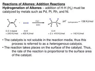
malonylCoA (4C) AcetylCoA carboxylase ATP ADP • first reaction committed step in fatty acid synthesis + HCO3- fatty acids AcetylCoA (2C) • Also uses biotin as swinging arm between two active sites • reactions very similar to pyruvate carboxylase
HCO3- ATP biotin ADP malony-lCoA (3C) biotin’s swinging arm carboxyphosphate Acetyl-CoA WOW look! mechanism of carboxylation (addition of COO-) is the same as for pyruvate carboxylase!!! ATP-dependent carboxylation of the biotin, carried out at active site 1 , is followed by transfer of the carboxyl group to acetyl-CoA at a second active site 2 . only difference is COO- is added to acetylCoA rather than to pyruvate (2C) carboxybiotin
regulation of AcCoA-Carboxylase The mammalian enzyme is regulated, by • phosphorylation by cAMP dependent kinase • inhibition when energy (cAMP) • allosteric control by local metabolites. Conformational changes with regulation: • active = multimeric filamentous complexes. • inactive = dissociation to = monomeric form P
fatty acid synthase • dimer • 6 active sites are individual domains of a large protein • ? developed from gene fusion • has more catalytic activities than any enzyme! • has two prosthetic groups thioester bonds • thiol of cysteine (in condensing domain) • thiol of P-pantetheine (in acyl carrier domain) • acts as a long flexible arm transferring substrates between active sites
has two prosthetic groups • thiol of cysteine in condensing domain • thiol of P-pantetheine Phosphopantetheine is covalently linked to a serine of the acyl carrier protein domain Thelong flexible arm of phosphopantetheine allows its thiol to move between active sites forms thioesters like CoA does
fatty acid synthase H20 2NADPH 3 2 2)Thioester bond between malonyl and pantetheine 3)The condensation reaction * involves decarboxylation of the malonyl carbanion attacks carbonyl carbon of the acetyl. Uses swinging arm of pantotheine You will have done these reactions in Dr Denyer’s lectures