Download

1 / 140
1.4k likes | 1.64k Views
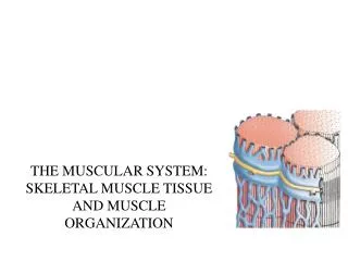
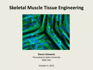
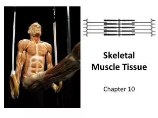
Skeletal Muscle Tissue. Skeletal Muscle Tissue Arrangement. Myofibrils – contractile elements of muscle tissue. Skeletal Muscle Cont. Muscle fiber – Muscle cell; composed of several myofibrils. Skeletal Muscle Cont.

E N D
Skeletal Muscle Tissue Arrangement • Myofibrils – contractile elements of muscle tissue
Skeletal Muscle Cont. • Muscle fiber – Muscle cell; composed of several myofibrils
Skeletal Muscle Cont. • Each muscle fiber is surrounded by a thin sheath of areolar connective tissue called endomysium
Muscle Tissue Cont. • Fascicles – A bundle of muscle fibers. There are usually between 10 to 100 muscle fibers in a fascicle.
Muscle Tissue Cont. • Each fascicle is surrounded by a layer of dense irregular connective tissue called perimysium
Muscle Tissue Cont. • Whole muscle – made up of several fascicles
Muscle Tissue Cont. • The whole muscle is surrounded by a dense irregular connective tissue called epimysium
Muscle Tissue • All three connective tissues (endomysium, perimysium, epimysium) extend beyond the muscle fiber to form a tendon.
Muscle Tissue • Tendon – Composed of dense regular connective tissue that attaches muscle to the periosteum of the bone
General Features of Skeletal Muscle • Striated
General Features of Skeletal Muscle • Voluntary
General Features of Skeletal Muscle • Multinucleated
General Features of Skeletal Muscle • Controlled by the somatic (voluntary) division of the nervous system
Microscopic Anatomy of Muscle Fibers • Muscle Fiber = Muscle Cell
Microscopic Anatomy cont. • Sarcolema – plasma membrane of muscle cells or muscle fibers
Microscopic Anatomy cont. • The multiple nuclei of each muscle fiber is located beneath the sarcolema
Microscopic Anatomy cont. • T (tranverse tubules) – Invagination of the sarcolema that tunnel in from the surface to the center of each muscle fiber
Microscopic Anatomy cont. • Sarcoplasm – cytoplasm of a muscle fiber
Microscopic Anatomy cont. • Sarcoplasmic reticulum – fluid filled system of membranous sacs. Calcium is stored here.
Microscopic Anatomy cont. • Dilated ends of SR are called terminal cisterns
Microscopic Anatomy cont. • Myofibrils are composed of functional units called sarcomeresresponsible for the striations
Microscopic Anatomy cont. • Each sarcomere is separated from the next by z discs
Microscopic Anatomy cont. • Sarcomeres are composed of thick (myosin) and thin (actin) filaments
Microscopic anatomy cont. • A band is the part of the sarcomere composed of thick (myosin) and thin (actin) filaments
Microscopic anatomy cont. • The A band is the dark striation seen under the microscope
Microscopic Anatomy cont. • I Band is the part of the sarcomere that contains only thin (actin) filaments
Microscopic Anatomy cont. • I Band is the light striation seen underneath the microscope
Microscopic Anatomy • The H zone is the part of the A band that contains only thick filaments (myosin)