Structure and Function of the Cardiovascular System
570 likes | 592 Views
Learn about the essential structures and functions of the cardiovascular system, including the heart, blood vessels, and circulation pathways. Understand how the system ensures oxygen and nutrient supply to the body's tissues and removes waste products. Discover the cardiac cycle and the importance of cardiac conduction for proper heart function.

Structure and Function of the Cardiovascular System
E N D
Presentation Transcript
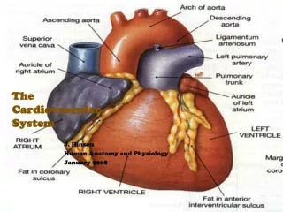
The CardiovascularSystem J. Hinson Human Anatomy and Physiology January 2008
A functional cardiovascular system is vital for survival because without circulation, the tissues lack a supply of oxygen and nutrients and waste substances begin to accumulate.
I. Structure of the Heart • Muscular pump w/in thoracic cavity superior to the diaphragm 1. w/in mediastinum; borders: laterally-lungs, posteriorly-spine, anteriorly-sternum • Size: 14 cm x 9 cm (slightly larger than the fist) http://butler.cc.tut.fi/~malmivuo/bem/bembook/06/fi/0601.gif
I. Structure of the Heart C. Base beneath 2nd rib; apex at 5th intercostal space D. Enclosed w/in parietal and visceral pericardium for some protection E. Three Layers 1. Epicardium (outer pericardium) 2. Myocardium (middle cardiac muscle) 3. Endocardium (inner epithelium); contains Purkinje fibers
http://www.cayuga-cc.edu/people/facultypages/greer/biol204/heart1/IMG00006.gifhttp://www.cayuga-cc.edu/people/facultypages/greer/biol204/heart1/IMG00006.gif
Structure of the Heart F. Chambers and Valves 1. Four Chambers a. Atria: upper/smaller b. Ventricles: lower/larger c. Septum divides R and L sides http://www.patienthealthinternational.com/sites/35/imagebank/typeArticleparam519594/heart_2.jpg
I. Structure of the Heart F. 2. Valves prevent blood flow in the opposite direction a. AV valve cusps connect to papillary muscles by chordae tendinae (i) Tricuspid and Bicuspid (mitral) b. 2 Semilunar (i) Pulmonary and Aortic http://butler.cc.tut.fi/~malmivuo/bem/bembook/06/fi/0601.gif
I. Structure of the HeartG. Blood Supply • Heart tissue receives nutrients from the coronary arteries. • Drained by the cardiac veins which joins the coronary sinus and empties into the RA. http://www.clevelandclinic.org/heartcenter/images/guide/heartworks/heartfront.jpg
II. Pathway of Blood through the Heart • Deoxygenated blood enters the RA. • From RA through Tricuspid Valve into RV. • From RV through Pulmonary Valve into Pulmonary Trunk. • From trunk into R and L Pulmonary Arteries to lungs for gas exchange. • Oxygenated blood from lungs to LA. • From LA through bicuspid (mitral) valve into the LV • From LV through Aortic Valve into the Aorta • From aorta to body and heart muscle.
II. Pathway of Blood through the Heart http://www.youtube.com/watch?v=PgI80Ue-AMo http://images.google.com/imgres?imgurl=http://library.thinkquest.org/25896/images/heart/real_animation.gif&imgrefurl=http://library.thinkquest.org/25896/sub_heart/action.htm&h=301&w=226&sz=35&hl=en&start=6&um=1&tbnid=KR4_0rNqoXoF3M:&tbnh=116&tbnw=87&prev=/images%3Fq%3Dblood%2Bflow%2Banimation%26svnum%3D10%26um%3D1%26hl%3Den%26rls%3Dcom.microsoft:en-us:IE-SearchBox%26rlz%3D1I7ADBS%26sa%3DN
http://images.main.uab.edu/healthsys/ei_0329.gif http://images.main.uab.edu/healthsys/ei_0329.gif
III. Pathways of Circulation • Pulmonary Circuit • From the heart to the lungs and back • From right ventricle enters pulmonary trunk and divides into R and L pulmonary arteries which travel to lungs (i) Only arteries carrying deoxygenated blood. • From lungs enters pulmonary veins and returns to left atrium (i) Only veins carrying oxygenated blood.
III. Pathways of Circulation http://training.seer.cancer.gov/module_anatomy/unit7_3_cardvasc_blood3_pathways.html
III. Pathways of Circulation B.Systemic Circuit • From the heart to the body and back • From LV oxygenated blood forced into the aorta (i) Thus, left heart muscle is larger! • Veins return via inferior and superior vena cava, then to right atria http://training.seer.cancer.gov/module_anatomy/unit7_3_cardvasc_blood3_pathways.html
III. Pathways of Circulation http://10.40.100.205/?a=175017&d=25355AA http://www.biosbcc.net/doohan/sample/images/heart/0283circulation.jpg
IV. Cardiac Cycle:atria contracted/ventricles relaxed; atria relaxed/ventricles contracted http://images.google.com/imgres?imgurl=http://acousticheart.com/library/HeartBeatingAnimation.gif&imgrefurl=http://acousticheart.com/learning_heart_and_lung_sounds.html&h=155&w=150&sz=59&hl=en&start=12&um=1&tbnid=Tl1JJcEOsG53GM:&tbnh=97&tbnw=94&prev=/images%3Fq%3Dcardiac%2Banimation%26svnum%3D10%26um%3D1%26hl%3Den%26rls%3Dcom.microsoft:en-us:IE-SearchBox%26rlz%3D1I7ADBS%26sa%3DN
IV. Cardiac Cycle • Heart Sounds • “lub-dup” • “lub” – ventricular contraction – AV valves close • “dup” – ventricular relaxation – semilunar valves close 2. Blood vibrates • Contraction as a unit • Functional syncytium • Sounds may identify heart conditions. Ex: murmur, alteration of valves, endocarditis http://www.youtube.com/watch?v=xS3jX1FYG-M&feature=related
V. Cardiac Conduction • The cardiac conduction system functions to coordinate the events occurring during the cardiac cycle. • Sinoatrial Node (S-A node) – primary • Specialized tissue at posterior RA • Cells excite themselves • Initiates 70-80 impulses per minute • “Pacemaker” • Atrioventricular Node (A-V node) – secondary • Located on floor of the RA near septum • May assume primary function if SA fails
V. Cardiac Conduction • Bundle of His: AV bundle at “crossroads” of septum • Purkinje fibers: spread throughout septum, walls, and to apex • Artificial pacemakers may treat disorders 1. Creates electrical pulse http://butler.cc.tut.fi/~malmivuo/bem/bembook/06/fi/0601.gif
V. Cardiac Conduction http://images.google.com/imgres?imgurl=http://www.arrhythmia.org/general/whatis/gifs/normal.gif&imgrefurl=http://defiant.corban.edu/jjohnson/Pages/HumPhys/Links/12CardioLinks.html&h=342&w=322&sz=50&hl=en&start=3&um=1&tbnid=DvDPrPdH-obNnM:&tbnh=120&tbnw=113&prev=/images%3Fq%3Dcardiac%2Banimation%26svnum%3D10%26um%3D1%26hl%3Den%26rls%3Dcom.microsoft:en-us:IE-SearchBox%26rlz%3D1I7ADBS%26sa%3DN
Electrocardiagrams (ECG) • Recording of electrical changes that occur in the myocardium during a cardiac cycle • Result of depolarization and repolarization associated with action potentials of cardiac muscle fibers http://video.about.com/heartdisease/Electrocardiogram.htm
Electrocardiagrams (ECG) • Readings: • P-wave: atrial depolarization • QRS complex: ventricular depolarization (masks atrial repolarization) • T-wave: ventricular repolarization • Time from P to Q [P-R interval] is the time it takes for an impulse to conduct from the SA to AV node
http://butler.cc.tut.fi/~malmivuo/bem/bembook/06/fi/0601.gif
VI. Blood Vessels • Arteries • Carry blood AWAY from the heart • Three layers • Tunica intima: innermost endothelium • Tunica media: middle smooth muscle • vasoconstriction: bv contract and diameter decreases • Vasodilation: bv relax and diameter increases • Tunica adventitia: outer connective tissue
VI. Blood Vessels • Arterioles • Continuations of arteries that join capillaries • Layers become progressively thinner • Capillaries • Smallest bvs; connect arterioles to venules • Single layer of squamous epithelium (endothelium) • Site of gas, nutrient, and waste exchange by diffusion, filtration, and osmosis • Fluid can enter causing edema.
VI. Blood Vessels • Venules • Connect capillaries to veins • Veins • Three layers with thinner walls and less smooth muscle (than arteries) • Contain valves that aid in returning blood to the heart and prevent backflow of blood
http://www.unm.edu/~jimmy/vessels.jpg http://www.unm.edu/~jimmy/vessels.jpg
VII. Blood Pressure • The pressure exerted by blood against the inner walls of the blood vessel • Most commonly aorta • Arterial blood pressure corresponds to cardiac cycle. • Systolic: maximum pressure during ventricular contraction • Diastolic: lowest pressure during ventricular relaxation • Written as systolic over diastolic: “normal” < 120/80 • Pulse: expand/recoil of arterial walls
VII. Blood PressureC. Influenced By: • Cardiac Output: vol. / minute • Stroke Volume: vol. / contraction • Formula: (i) “normal” SV=70 mL; “normal” HR=60-80 bpm (ii) SV and HR directly related to BP • Blood Volume: • 4-6 L • directly related to BP
VII. Blood PressureC. Influenced By: • Peripheral Resistance: friction • Vasoconstriction increases; Vasodilation decreases • PR is directly related to BP • Viscosity: ease of fluid movement • Directly proportional to BP
VII. Blood Pressure • Regulated by parasympathetic nervous system • Blood flow through the venous system is low; depends upon muscle contraction and respiratory movements ** Sphygmomanometer http://www.knowledgebase-script.com/demo/admin/attachments/blood-pressure-check.jpg http://static.howstuffworks.com/gif/adam/images/en/blood-pressure-picture.jpg
VII. Cardiovascular Disease J. Hinson Human Anatomy and Physiology January 2007
B. Statistics • #1 cause of death in US at 36.3% • coronary artery disease #1; stroke #3 • 1 person dies every 30 sec (~ 2600/day) • ~100,000 under age 50 • 71 million Americans suffer from disease • Costs US $274 billion dollars • (American Heart Association, 2007) • (http://www.healingwithnutrition.com)
http://www.healingwithnutrition.com/cdisease/cardiovascular/cardiovascular.html#A1http://www.healingwithnutrition.com/cdisease/cardiovascular/cardiovascular.html#A1
C. Risk Factors • GENETICS • Exercise • Diet • Smoking • Obesity • Stress • high cholesterol • high blood pressure • Diabetes
http://battlingforhealth.com/wp-content/uploads/2008/06/us-map.gifhttp://battlingforhealth.com/wp-content/uploads/2008/06/us-map.gif
D. Hypertension • High blood pressure • called “silent killer” as most have no symptoms • BP > 140/90 • 90-95% of unknown cause http://www.zyduscadila.com/hyperday/hype_images/hypertension_r34_c2.jpg
E. Congestive Heart Failure • heart is unable to pump an adequate amount of blood to the body cells • heart transplant candidates http://video.about.com/heartdisease/Congestive-Heart-Failure.htm
F. High Cholesterol • Characterized by: • Total cholesterol: > 200 mg/dL • Triglycerides: > 150 mg/dL • HDL (good): < 40 mg/dL • LDL (bad): > 100 mg/dL
G. Atherosclerosis • Fatty plaques clogs artery • Pieces may break away to become emboli, or clots • Tx: angioplasty • May lead to Coronary Artery Disease which can lead to… http://www.youtube.com/watch?v=N7nghr9TpSU
Atherosclerosis http://www.apsu.edu/thompsonj/Anatomy%20&%20Physiology/2020/2020%20Exam%20Reviews/Exam%201/atherosclerosis01a.bmp
Acute ischemic attack causing death of heart tissues Often involves the coronary artery Symptoms: Primary: Chest pain (angina pectoris) and tightness Other sxs: shortness of breath, nausea, sweating, radiating pain Treatment: nitroglycerin Relaxes veins to decrease stroke volume Relaxes arteries to increase blood flow H. Myocardial Infarction: Heart Attack
I. Sudden Cardiac Arrest • heart abruptly stops beating, usually associated with coronary artery disease http://www.youtube.com/watch?v=_3omQ_mKBPs
J. Cerebrovascular Accidents: STROKE • Result of decreases blood flow to the brain • 70-80% due to clot; or caused by hemorrhage • Sxs: inability to speak, confusion, loss of feeling/function on one side of the body, and severe headache • Transient Ischemic Attack (TIA): “mini-stroke” • no lasting affect; no apparent damage • often mimics migraines • Tx: Tissue Plasminogen Activator (TPA) w/in 6-8 hours to help dissolve clot


