Cell Theory
Cell Theory. Topic 2.1. Assessment Statements. 2.1.2 Outline the cell theory. 2.1.2 Discuss the evidence for the cell theory 2.1.3 State that unicellular organisms carry out all the functions of life

Cell Theory
E N D
Presentation Transcript
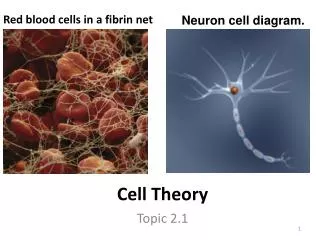
Cell Theory Topic 2.1
Assessment Statements 2.1.2 Outline the cell theory. 2.1.2 Discuss the evidence for the cell theory 2.1.3 State that unicellular organisms carry out all the functions of life 2.1.4 Compare the relative sizes of molecules, cell membrane thickness, viruses, bacteria, organelles and cells, using the appropriate SI unit 2.1.5 Calculate the linear magnification of drawings and the actual size of specimens in images of known magnification
2.1.6 Explain the importance of the surface area to volume ratio as a factor limiting cell size 2.1.7 State that multicellular organisms show emergent properties 2.1.8 Explain that cells in multicellular organisms differentiate to carry out specialized functions by expressing some of their genes but not others 2.1.9 State that stem cells retain the capacity to divide and have the ability to differentiate along different pathways 2.1.10 Outline one therapeutic use of stem cells
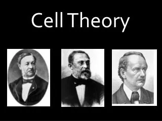
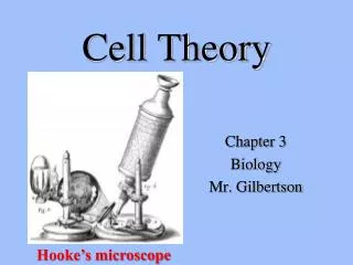
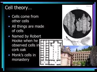
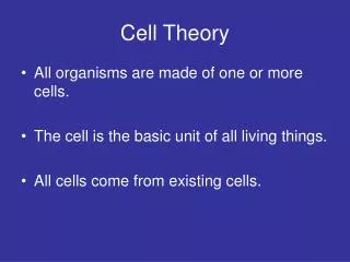
Cell theory • All organisms are composed of one or more cells • Cells 1st observed in 1665 by Robert Hooke while observing cork with a microscope he built • Antonie van Leeuwenhoek observed 1st living cells, “animacules” • In 1838 Mathias Schleiden stated that plants are made of “independent, separate beings” called cells • In 1839 Theodor Schwann made a similar statement about animals
Cells are the smallest units of life • No living entity found has not been made of at least one cell • All cells come from pre-existing cells • Supported by Louis Pasteur in 1860s through experiments that disproved spontaneous generation • Possible exceptions?
Metabolism (all chemical reactions that occur within an organism) Growth Reproduction (hereditary molecules that can be passed to offspring) Response Homeostasis (maintenance of a constant internal environments) Nutrition (providing a source of compounds that can be broken down to provide organism with energy and nutrients) Functions of life
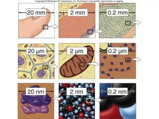
Cells and sizes • Most cells are up to 100 μm (micrometers) • Organelles are up to 10 μm • Bacteria are up to 1 μ • Viruses are up to 100 nm (nanometers) • Membranes are 10 nm thick • Molecules are near 1 nm • All objects are 3-D
Calculating size • Know the diameter of the microscope’s field of vision using a simple ruler • Size of specimen can then be calculated in the field • By knowing the diameter of the field of view and having an estimate of the number of cells that would fit across the diameter, you can determine the size of a cell by dividing the diameter by the number of cells.For example:The diameter of the field of view under 100 total magnification is about 1.5 mm. If there are 10 cells that would fit across the diameter, one cell would be 0.15mm.The diameter of the field of view under 400 total magnification is approximately 0.375 mm.
Light microscope type found in most schools (including ours), uses compound lenses and light to magnify objects The lenses bend, or refract the light, which makes the object beneath them appear closer. Magnifies objects up to 2000 times. Two dimensional images Poor resolution Bonuses: Relatively inexpensive, can view living organisms in color Electron microscope use electrons (negatively charged electrical particles) to view the specimen Types: Scanning 3D images Magnifies 50,000 X Non-living specimens Transmission 2D images Magnifies 2,000,000 X Non-living specimens High magnification and resolution Types of microscopes
Limiting cell size • Why must cells be small? • Surface area to volume ratio • Homeostasis dependent upon volume • Greater surface area able to move more materials in and out • A large cell has relatively less surface are than a small cell • What about large cells?
Calculating SA/V of a sphere • SA=4πr2 • V=(4/3) πr3
Cell reproduction and differentiation • Allows possibility of growth and replacement of damaged or dead cells • Multicellular organisms begin as a single cell that reproduces at a rapid rate • Resulting cells go through differentiation as a result of the expression of genes • Some cells lose ability to reproduce once they become specialized
Stem cells • What are stem cells? • Populations of cells that retain their ability to divide and differentiate into various cell types • Ex. Meristematic tissue in plants, embryonic (pluripotent) in animals • Cannot be distinguished by appearance; only by behavior
Stem cell research • Directed towards growing large numbers of embryonic stem cells in culture • Used to replace differentiated cells lost due to injury and disease • Parkinson’s disease and Alzheimer’s disease are caused by loss of brain cells and it is hoped that implanted stem cells could replace many of these lost brain cells • Other examples: diabetes, leukemia
Ethical issues • What is the controversy? • Where do you stand in the debate? • How do you feel about the source of pluripotent stem cells?
Prokaryotic Cells Topic 2.2
Assessment Statements 2.2.1 Draw and label a diagram of the ultrastructure of Escherichia coli as an example of a prokaryote 2.2.2 Annotate the diagram with the functions of each named structure 2.2.3 Identify structures from 2.2.1 in electron micrographs of E. coli 2.2.4 State that prokaryotic cells divide by binary fission
What is a prokaryotic cell? • Most are less than 1 μm in diameter • DNA not enclosed within a membrane and is one circular chromosome • DNA not attached to proteins • Lack membrane-bound organelles • Cell wall made of peptidoglycan • Divide by binary fission
E. Coli ultrastructure • Be able to draw and label: • Cell wall • Plasma membrane • Flagella • Ribosomes • Nucleoid (region containing free DNA)
Cell wall • Protects and maintains the shape of the cell • Composted of carbohydrate-protein complex called peptidoglycan • Some bacteria have an additional layer of polysaccharide (capsule) outside cell wall which makes it possible for some to adhere to structures
Plasma membrane • Just inside cell wall • Controls movement of materials in and out of the cell • Plays a role in binary fission • Cytoplasm occupies the interior of the cell and is location for all cellular processes • Most visible structure is single chromosome
Pili • Hair-like growths on outside of the cell wall • Used for attachment • Main function is joining bacterial cells in preparation for the transfer of DNA from one cell to another
Flagella • (sing.) or flagellum (pl.) • Longer than pili • Allow cell motility
Ribosomes • Occur in all prokaryotic cells • Function as sites of protein synthesis • Occur in very large numbers in cells with high protein production
Nucleoid region • Non-compartmentalized • Contains a single, long, continuous, circular thread of DNA • Involved in cell control and reproduction • Cell may also contain plasmids which replicate independently of the chromosomal DNA • Plasmids are not required but may help the cell adapt to unusual circumstances
Binary fission • Process by which prokaryotes divide • DNA is copied • 2 daughter chromosomes become attached to different regions on the plasma membrane • Their movement is aided by fibers made of protein called FtsZ • Cell divides into two genetically identical daughter cells
Paul the Prokaryote • While watching the clip list as many facts about prokaryotes that you see depicted
Eukaryotic Cells Topic 2.3
Assessment Statements 2.3.1 Draw and label a diagram of the ultrastructure of a liver cell as an example of an animal cell 2.3.2 Annotate the diagram with the functions of each named structure 2.3.3 Identify structures from 2.3.1 in electron micrographs of liver cells 2.3.4 Compare prokaryotic and eukaryotic cells 2.3.5 State three differences between plant and animal cells 2.3.6 Outline two roles of extracellular components
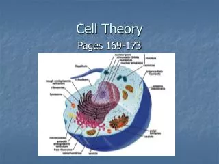
What is a eukaryotic cell? • Range in diameter from 5 to 100 μm • Noticeable nucleus • Compartmentalized due to presence of organelles (non-cellular structures which carry out specific functions)
Common organelles • Endoplasmic reticulum • Ribosomes • Lysosomes • Golgi apparatus • Mitochondria • Nucleus • Chloroplasts • Centrosomes • Vacuoles
Cytoplasm • Within plasma membrane • Fluid between organelles is called cytosol
Extensive network of tubules or channels Transports materials throughout the internal region of the cell Two types: Rough (has ribosomes attached) Smooth (lacks ribosomes) Rough Involved in protein development and transport Closer to nuclear membrane Smooth Produces lipids, sex hormones Detoxifies drugs Stores calcium ions Transports lipid-based compounds Aids liver in release of glucose Endoplasmic reticulum
Ribosomes • Carry out protein synthesis • May be free or attached to ER • Composed of RNA and protein • Larger and denser than those found in prokaryotes
Lysosomes • Digestive centers that arise from the Golgi apparatus • Contains enzymes that break down proteins, nucleic acids, lipids and carbohydrates
Golgi apparatus • Made of flattened sacs called cisternae • Collects, packages, modifies and distributes materials synthesized in the cell
Mitochondria • Produces usable cellular energy called ATP through process of cellular respiration • Has its own DNA and ribosomes • Capable of reproducing independent of cell
Nucleus • Region where DNA is located • Bordered by nuclear envelope which has numerous pores that allows communication with the cell’s cytoplasm • DNA occurs in form of chromosomes or chromatin • Some cells extrude their nucleus and are more specialized for a specific function • Nucleolus within nucleus produces ribosomes
Chloroplasts • Occur only in plant and algae cells • Contains its own DNA in the form of a ring • Includes grana, thylakoids, and stroma • Carries out photosynthesis • Capable of reproducing independent of cell
Centrosome • Pair of centrioles at right angles to one another • Involved in assembling microtubules which provide structure and movement of cell
Vacuoles • Formed from Golgi apparatus • Store potential food, metabolic wastes, toxins, and water • Enable plants to have higher surface area to volume ratios even at larger sizes • In plants, they provide rigidity when filled with water
Prokaryotic cells DNA in a ring w/out protein DNA free in cytoplasm No mitochondria 70S ribosomes No internal compartmentalization < 10 μm Eukaryotic cells DNA with proteins as chromosomes/chromatin DNA enclosed Mitochondria present 80S ribosomes Internal compartmentalization > 10 μm Prokaryotic vs. Eukaryotic
Prokaryotic AND Eukaryotic • Both have outer boundaries that always involves a plasma membrane • Both carry out all the functions of life • Both have DNA
Plant cells Exterior of cell includes cell wall Chloroplasts present Large central vacuole Store carb. as starch Do not contain centrioles Has a fixed, often angular shape Animal cells Only plasma membrane; no cell wall No chloroplasts Vacuoles not present or small Store carb. as glycogen Contain centrioles Cell is flexible and more likely to be rounded in shape Plant vs. Animal
Bacteria Cell wall of peptidoglycan Fungi Cell wall of chitin Yeasts Cell wall of glucan and mannan Algae Cell wall of cellulose Plants Cell wall of cellulose Animals No cell wall Extracellular matrix made of glycoproteins Outermost regions
Cell wall maintains cell shape Helps regulate water intake Extracellular matrix (ECM) Composed of collagen and glycoproteins Strengthens plasma membrane Allows for cell-to-cell interaction, possibly altering gene expression Directs stem cells to replicate Cell migration and movement Functions of extracellular components