SYNAPSE:
SYNAPSE:. Dr. Ayisha Qureshi MBBS, Mphil Department of Physiology. Synapse:. Definition : A synapse is a region of functional contact and anatomical differentiation between two neurons. OR It is a point of contact between two adjacent neurons.


SYNAPSE:
E N D
Presentation Transcript
SYNAPSE: Dr.AyishaQureshi MBBS, Mphil Department of Physiology
Synapse: Definition: A synapse is a region of functional contact and anatomical differentiation between two neurons. OR It is a point of contact between two adjacent neurons. • Action potentials cannot cross the synaptic cleft present between 2 neurons. • Nerve impulse is carried by neurotransmitters which transmit the nerve impulse from one nerve cell to the next across the synapse. • The structure of synapse consists of: • presynaptic ending (from where neurotransmitters in vesicles are synthesized & released) • post synaptic ending (has neuroreceptors in the membrane) • synaptic cleft
Types of Synapses: • Chemical Synapse (transmission thru chemicals i.e. NT) • Electrical Synapse • Impulse conducted without release of NT • Synaptic gap only 2-3 nm • No synaptic delay • Unidirectional & Bidirectional conduction • Mixed Synapse i.e. having both electrical & chemical regions
CLASSIFICATION OF SYNAPSES: Anatomical classification of Synapses: • Axo-dendritic • Axo-somatic • Axo-axonic • Somato-dendritic • Dendro-dendritic • Somato-somatic • Reciprocal • Serial • Triad
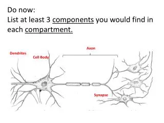
Structure Of a Synapse SYNAPSE= Presynaptic terminal + Synaptic cleft + Postsynaptic terminal • Presynaptic terminal: is the first part of the synapse & is usually (not always) the Axon terminal. The axon terminals are also called the boutonterminaux or synaptic knob. The synaptic knobs have synaptic vesicles that contain the NT (neurotransmitters). The NT are produced in the body & conducted along the axon (anterograde flow). The NT can be inhibitory or excitatory. • Synaptic cleft or gap:is app. 20nm. It is a non-anatomical continuity between the post and pre-synaptic ends. • Postsynaptic terminal: is the name given to the last part of the synapse. It is usually comprised of the dendrite or the cell body on which the axon synapses.
Mechanism Of Conduction of an Impulse in a chemical synapse • action potential reaches the PRESYNAPTIC terminal ↓ • voltage-gated Ca2+ channels open ↓ • influx of Ca2+ ↓ • synaptic vesicles fuse with the pre-synaptic membrane (exocytosis) ↓ • neurotransmitters are released into SYNAPTIC TERMINAL cross it and diffuse to the POST-SYNAPTIC terminal ↓ • neurotransmitter binds to neuroreceptor on postsynaptic membrane ↓ • causes Na+ channels to open, and Na+ flows into postsynaptic membrane ↓ • if threshold is reached then action potential is initiated ↓ • neurotransmitter is broken down by specific enzymes in the synaptic cleft.
Fate of the Neurotransmitter: Dissociates from the Receptor & can have either of the 3 fates: • Enzymatic Degradation: A portion of it is inactivated by the enzymes present in high concentration at the postsynaptic membrane. • Re-uptake of remaining NT by Pre-synaptic neuron and Re-used. • Diffusion into the blood stream.
GRADED POTENTIAL • Let’s consider a stimulus at the dendrite of a neuron. The stimulus reaches the dendrite (postsynaptic neuron) from the axon (presynaptic neuron) with the help of a NT. • The NT leads to opening of simple ligand-gated channelsthat are present in the postsynaptic membrane, either Na+ or K+ channels which leads to Na or K or Influx; this could lead to depolarization or repolarization. • However, dendrites and somata typically lack voltage-gated channels, which are found in abundance on the axon hillock and axolemma. They only contain Na channels that depend upon the NT…. • So what cannot occur on dendrites and somata? An Action Potential cannot occur in the soma & dendrites
Thus, the question we must answer is, “what does the depolarization that results due to the opening of the ligand gated Na channels do?” This depolarization leads to the generation of a aGRADED POTENTIAL….
Graded Potentials • The positive charge carried by the Na+ spreads as a wave of depolarization through the cytoplasm (much like the ripples created by a stone tossed into a pond). • As the Na+ drifts, some of it will be moved back into the ECF by the NA-K pump. • What this means is that the degree of depolarization caused by the graded potential decreases with distance from the origin unlike the AP.
Graded Potentials (GP) • Their initial amplitude may be of almost any size – it simply depends on how much Na+ originally entered the cell, which depends on how many NT molecules were released. • If the initial amplitude of the GP is sufficient, it will spread all the way to the axon hillock where Voltage-gated Na channels reside. • If the arriving potential change is threshold or suprathreshold, an ACTION POTENTIAL will be initiated in the axon hillock and it will travel down the axon. If the potential change is subthreshold, then NO AP will initiated and NOTHING will happen.
The kind of ion that enters the Postsynaptic terminal (whether Na or K) will determine the kind of potential that will be generated: Depolarization OR Repolarization.
EPSP (Excitatory postsynaptic potential) IPSP (Inhibitory postsynaptic potential) Opening of Chloride channels & chloride ion influx Increased efflux of K ions Both cause increased negativity inside the neuron leading to repolarization and inhibition of the neuron. Thus, AP will NOT be initiated… • Opening of Na channels • K & cl channels are not opened. Both the above actions cause increased positivity of the neuron and so excitation that can lead to depolarization & AP….
GRADED POTENTIAL EPSP IPSP
1. DALE’S LAW: This law states: At a given chemical synapse only one type of neurotransmitter is released and thus only one effect, either excitatory or inhibitory, is possible.
2. IRREDUCIBLE SYNAPTIC DELAY Definition: It is the time taken for the neurotransmitter to be released from the presynaptic membrane, diffuse across the synaptic cleft to reach the post synaptic membrane and bind to the neuroreceptors there. It is about 0.5 msec.
3. ONE-WAY TRAVEL In a chemical synapse the impulse always travels from the presynaptic to the postsynaptic cell as the neurotransmitter is only released from the presynaptic terminal.
4. CONVERGENCE Usually the postsynaptic neuron receives afferents from a large no. This means that a number of neurons will synapse on a single neuron. It is very rare to find that only a single neuron synapses on another single neuron. 1:1 convergence is rare.
5. SPATIAL SUMMATION Summation of stimuli from two different presynaptic elements reaching a neuron simultaneously, which by adding up results in excitation or facilitation, of a postsynaptic neuron is called SPATIAL SUMMATION.
6. TEMPORAL SUMMATION Summation of stimuli from two different presynaptic impulses reaching a neuron one after the other, which by adding up results in the excitation or facilitation of a postsynaptic neuron is called TEMPORAL SUMMATION.
7. EFFECTS OF CHEMICAL CHANGES IN THE BLOOD: • Acidosis depresses while alkalosis increases the neuronal activity. • Hypoxia exerts a depressing effect.
8. OCCLUSION: The deficit in the effect of the discharge of the two preganglionic neurons on being simultaneously stimulated is called OCCLUSION. (Figure: 2-20, page: 136, Essentials of Medical Physiology by Mushtaq)
9. INHIBITION: • Feed- Forward Inhibition • Presynaptic Inhibition • Recurrent Inhibition (Figure: 2-21, page: 137, Essentials of Medical Physiology by Mushtaq, 5th edition)
10. FATIGUE If there is continuous stimulation of the presynaptic synapse, this leads to the neurotransmitter supply being exhausted. This causes the synaptic transmission to stop.