Download
1 / 16
160 likes | 253 Views
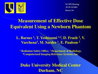
NC HPS Meeting 10/18-19/2001 Boone, NC. Measurement of Effective Dose Equivalent Using a Newborn Phantom L. Barnes 1 , T. Yoshizumi 1,2 , D. Frush 2 , V. Varchena 3 , M. Sarder 1 , E. Paulson 2 1 Radiation Safety Office, 2 Department of Radiology,

E N D
NC HPS Meeting10/18-19/2001Boone, NC Measurement of Effective Dose Equivalent Using a Newborn Phantom L. Barnes 1, T. Yoshizumi 1,2, D. Frush 2, V. Varchena3, M. Sarder 1, E. Paulson 2 1 Radiation Safety Office, 2 Department of Radiology, 3Computerized Imaging Reference Systems, Inc. Duke University Medical Center Durham, NC
Measurement of Effective Dose Equivalent Using a Newborn Phantom Topics • Why pediatric CT dosimetry? • Scope of study • Materials and Methods • Results • Conclusions
Why pediatric CT dosimetry? • Only 40% of CT users adjusttechniques for patient size (preliminary NEXT data) • NEXT =Committee on Nationwide Evaluation of X-ray Trend, CRCPD • Don’t have organ dose data in multi-detector CT scanners (your guess is as good as mine) • Dose indices such as CTDI and the dose-length product do not represent actual organ dose and are of limited value in risk assessment • Problems created by news media frenzy in recent months
2. Scope of study • Measure Effective Dose Equivalent using single and multi-detector CT scanners for chest and abdomen CT protocols; • Two protocols were selected: Chest and Abdomen; • Scan parameters (kVp, mA, sec, pitch, etc.) were selected to represent High, Medium, and Low techniques.
3. Materials and Methods • Dosimeters • Harshaw TLD-100 • Harshaw auto TLD reader QS 5500 • CT scanners • GE QXi (multi-detector) and CTi (single detector) • Anthropomorphic phantom • Newborn phantom, CIRS, Inc., Norfolk, VA.
Brief description of phantom • Atom newborn phantom (Model 703-D) CIRS, Norfolk, VA • Cost: ~ $ 9K • Joint effort between Duke and CIRS
Dosimeter distribution • TLD locations in organs pre-drilled • Designed for TLD-100 (3mm x 3 mm x 1 mm)
CTI High 3 mm, pitch 1.0 140 kVp;120 mA, 0.8 sec Medium 5 mm, pitch 1.5 140 kVp; 90 mA; 0.8 sec Low 5 mm, pitch 2.0 120 kVp; 70 mA; 0.8 sec QXI High 2.5/7.5 HQ 140 kVp; 100 mA, 0.8 sec Medium 3.75/11.25 HQ 140 kVp; 70 mA, 0.8 sec Low 5.0/22.5 HS 120 kVp; 60 mA, 0.5 sec Newborn Abdomen CT ProtocolDose Comparison: CT/i vs QX/i
ICRP Report No. 26 (1977) Effective Dose Equivalent = ST WT HT Where WT = weighting factor; HT = dose equivalent. Selected Organs (Newborn Phantom – CIRS, Norfolk, VA) –see Chart (Rt). Calculation of Effective Dose Equivalent
CTI High 3 mm, pitch 1.0 140 kVp;100 mA, 0.8 sec Low 5 mm, pitch 2.0 120 kVp; 50 mA; 0.8 sec QXI Plus High 2.5/7.5 HQ, 140 kVp, 80 mA, 0.8 sec Med 3.75/1.25 HQ, 140 kVp, 50 mA, 0.8 sec Low 5.0/22.5 HS 120 kVp; 40 mA, 0.5 sec Newborn Chest CT ProtocolDose Comparison: CT/i vs QX/i plus
ICRP Report No. 26 (1977) Effective Dose Equivalent = ST WT HT Where WT = weighting factor; HT = dose equivalent. Selected Organs (Newborn Phantom – CIRS, Norfolk, VA) –see Chart (Rt). Calculation of Effective Dose Equivalent
5. Conclusions • For abdomen protocol, the effective dose equivalent between high and low scan techniques differed a factor of 7 for QXi and that of 5 for CTi. • For chest protocol, the effective dose equivalent between high and low scan techniques differed a factor of 6 for QXi and 8 for CTi. • It is important to adjust scan techniques for the size and weight of a patient. • A multi-detector scanner (QXi) resulted in substantially higher dose than a single-detector scanner (CTi).