Download
1 / 32
510 likes | 1.67k Views
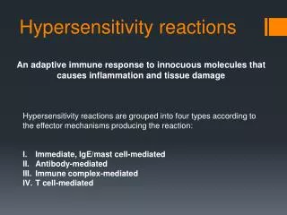
Hypersensitivity Reactions. Immunology Unit Department of Pathology College of Medicine King Saud University. Objectives. To know that hypersensitivity reactions are over and excessive immune responses that can be harmful to body in four different ways

E N D
Hypersensitivity Reactions Immunology Unit Department of Pathology College of Medicine King Saud University
Objectives • To know that hypersensitivity reactions are over and excessive immune responses that can be harmful to body in four different ways • To be familiar with inflammatory processes in Type I hypersensitivity reaction that mediate allergic inflammation • Recognize that Type II hypersensitivity deals with immune responses against antigens that are integral part of cell membrane and are usually associated with autoimmune disorders • To know that Type III hypersensitivity reactions are mediated by immune complexes and cause vasculitis • Describe Type IV hypersensitivity is a purely cell mediated immune response associated with chronic inflammation
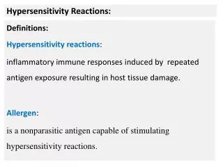
What is hypersensitivity? • Protectiveimmunity: desirable reaction • Hypersensitivity: undesirable reaction • Undesirable responses can be mediated by • Antibody binding to antigens (Types I-III) • Cell mediated reaction to chemicals or proteins (Type IV)
Type IV: Cell Mediated Immunity Type I: IgEAb Type II: IgGAb to tissue antigens Type III: IgGImmune Complexes Gel and Coombs Classification
Type I: Immediate Hypersensitivity • Most people will not react to these allergens but some individuals “atopic” respond by producing large amounts of IgE • Non-allergicindividuals respond to these allergens by producingIgGantibodies
Type I Hypersensitivity • Also termed as: Immediate Hypersensitivity Anaphylactic reactions Allergic reactions (Occurs within minutes to hours)
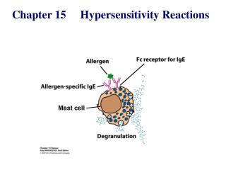
Features • Antibody type: IgE - Cellular components: Mast cells, basophiles & eosinophils - Antigens: Also known as allergens (antigens with low molecular weight & highly soluble)
Allergens • Some of the allergens involved in type I hypersensitivity are: pollens, dust mite allergens, animal dander, nuts, shellfish, various drugs etc
Type I Reaction occur in 2 phases: TypeIreactions occurin two phases • - Sensitizationphase • First contact with allergens - Challenge phase Subsequent contact with allergens • Phase I : - Sensitization phase . Allergen enter tissues , induce an immune response . B – cells transform to plasma cells & produce IgE. - IgE bind to receptors on Mast cells and basophiles ( F c ЄRI - high affinity receptors). individuals become : “ Sensitized . “ • Phase I : - Sensitization phase . Allergen enter tissues , induce an immune response . B – cells transform to plasma cells & produce IgE. - IgE bind to receptors on Mast cells and basophiles ( F c ЄRI - high affinity receptors). individuals become : “ Sensitized . “
Type I Hypersensitivity (Immediate) Sensitization Challenge
Allergy is a systemic disorder Nose Pharynx Allergic rhinitis Asthma Lungs Esophagus Food allergy Stomach Skin Eczema Urticaria Allergic dermatitis
Normal nose Allergy: Rhinitis, Eczema & Conjunctivitis
* Injected allergens: Bee sting venom enters the blood stream Systemic inflammation Anaphylactic shock (life - threatening) • Anaphylactoidreactions:- Are non - IgEmediated may result from contrast media or local anesthetics
Diagnosis of Allergy Skin Prick test 1. Skin prick test (SPT) 2. Specific IgE measurement (RAST) 3. Elimination / Provocation test (Food allergy)
Type II Hypersensitivity Reactions • Features:- • - IgG (or IgM) • - Antigens: bound to cell membranes • (Self antigens) - Exogenous antigens • (microbial) • - Complement activation (Invariable)
Clinical examples: Glomerulonephritis (anti-glomerular basement membrane) Mis-matched blood transfusion (the target ??)
Diagnosis - Detection of antibodies and antigens by Immunofluoresence in tissue biopsy specimens e.g. kidney, skin etc.
Type III: Immune complex hypersensitivity • When an antigen reacts with an antibody the product they form is called an immune complex which is capable of inducing an inflammatory response • Immune complexes are deposited in tissues like kidneys (nephritis), joints (arthritis) or blood vessels (vasculitis)
Type III Hypersensitivity(immune–complex mediated) • Features Antibody (IgG/ or IgM) + Antigen (soluble) - Immune – Complex formation - Complement activation - Attraction of inflammatory cells
Type III H/S: Clinical examples Glomerulonephritis (Blood vessel wall) Rheumatoid Arthritis , SLE
Diagnosis (Type III H/S) Demonstration of specific immune complexes in the blood or tissues by: Immunofluoresence
Type IV hypersensitivity reactions(Delayed Hypersensitivity) • Features • Cell mediated immune response • Antigen dependent T cell (CD4 generally and CD8 occasionally) activation via MHC Class I or II • Activated macrophages • Delayed onset (2-4 days) • Abnormal cellular response • (Granuloma formation)
Development of DTH Response Sensitization phase: 1-2 week period Effector phase: 24-72 hours Effector cells (activated macs) act non-specifically
Type IV clinical examples: Allergic contact dermatitis TB granuloma (persistent antigen)
Diagnosis (Type IV) 1. Delayed skin test (Mantoux test) 2. Patch test (Contact dermatitis) 3. Lymphocyte transformation test
Take Home Message • 1. Type I (IgE), II (IgG) and III (IgG) hypersensitivity reactions are mediated by antibodies whereas Type IV hypersensitivity reaction is a cell mediated immune response. • 2. Hypersensitivity reactions are undesirable, excessive, and aberrant immune responses associated with disorders such as allergy, autoimmunity and chronic inflammation.