Download
1 / 30
300 likes | 394 Views
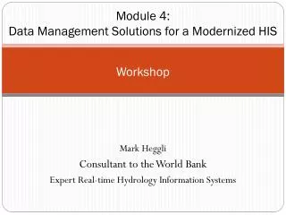
EBBEP Workshop. FP Plasmid Digest Lab. Where the FP came from. There are 6 different FP used for this lab available through EBBEP. Of the 6 proteins; 3 are from the Aequorea victoria jellyfish and 3 are from Discosoma striata anemone. Aiquorea victoria jelly. Discosoma striata anemone.

E N D
EBBEP Workshop FP Plasmid Digest Lab
Where the FP came from There are 6 different FP used for this lab available through EBBEP Of the 6 proteins; 3 are from the Aequorea victoria jellyfish and 3 are from Discosoma striata anemone Aiquorea victoria jelly Discosoma striata anemone
Structure of FP proteins All of the proteins used by EBBEP have a similar structure.They are all monomer barrel shaped molecules with a central chromatophore Small changes in the Amino acids in or directly Around the chromatophore cause changes in color
All proteins were made by Mutagenesis of the natural occurring proteins found in types of Cnidarians. How were the proteins made? Proteins made from anemone RFP Tangerine (orange/pink) Cherry (Dark pink) Grape (purple) Proteins made from Jellyfish GFP Emerald (Green) Venus (yellow) Blue
Using evolution • Since the proteins come from two different “roots” of evolution you can see the inheritance in the sequences of the proteins
The FP plasmids • Made from pRSET vector • FP gene is less than 730bp • Only difference in plasmids is the FP gene
Restriction enzymes • Type 2 restriction endonucleases are used in this lab. (most common) • Cut DNA at a specific sequence. • Enzymes used • HindIII • AhdI
Evolved by bacteria to protect against viral infection Over 3000 known enzymes Why are there restriction enzymes?
Challenges in making the lab • Needed an enzyme that cut green based genes different than red based genes. • That’s easy! Several to choose from • Finding enzymes that cut the plasmid into a reasonable number of bands and have bands of an easily visible size on a gel • Not so easy
Plasmid maps • Provided are 2 maps (red based and green based)
Reading the maps and predicting size • HindIII only cuts the plasmid once making 1 linier piece the full size of the plasmid ~3623bp
Reading the maps and predicting size AhdI cuts 2 times producing 2 fragments Size of fragment 1 2555-445= 2100bp Size of fragment 2 has 2 way to calculate 3623-2110 = 1513bp Or 3623-2555+445= 1513
Teacher set-up tips • Get labeling tubes early! • DNA, Buffers, Water, loading dye, and marker/ ladder are directly aliquotted from stocks supplied • Can be completed a few days in advance lf you have a freezer. More if you have a frosted freezer
Teacher Set-Up tips • Enzymes must be diluted. • Receive a stock enzyme tube and a dilution tube for each enzyme. • Add contents of one tube to the other (pre measured for you) • Must be done no earlier than 24 hrs before or enzymes will not work as well
Teacher set up tips • Sterile 1.5 snap cap tubes and filter pipette tips are ideal. • Will work without the Sterile but you will possibly see fuzzier bands in the gel. • Cap tubes before you give them out to help decrease the contamination
Teacher Set-Up tips • Make sure you plan for a long day. Staining gels with EtBr takes time. • If you don’t have a digital imaging system then remember you digital camera.
Things to stress with students: • Set up Digests on ICE! • Concentrations are important (More is not better) • Add reagents in Correct order. • Water first ,Buffer, DNA, Enzymes are always last. • Reason for Enzymes being last: Enzymes are sensitive to conditions outside of their normal range. Strong buffer solutions can effect the efficiency of the digest
Lab Procedure • Each group of 2 students sets up 3 tubes. • Negative control (no enzyme) • An undigested plasmid shows as usually 2 or more bands (nicked/open circle, super super coiled,multimers Travels Faster Travels Slower bs.kaist.ac.kr
Lab procedure • HindIII digest-Should make 1 linier fragment. Shows true size of plasmid • AhdI digest- determines if plasmids are from red (2 bands) or green (1band) line. • 2 Groups per gel with marker in middle. • Best to give 1 green and 1 red to each gel.
Lab procedure • Incubate at 37C. • Freeze over night (fridge will work for 24hrs) • Run gel. The bands are far enough apart you can run it pretty fast… if you can live with the smile bands on the gel
Keep it clean and don’t digest too long • Contaminations of DNases can be devastating. Lane 8 has little to none DNase activity Lane 1 has a large amount of DNase activity biosyn.com
Things that can go wrong • Impeded digestion due to incorrect set up. (too much or too little buffer etc.) • Star digest activity • Under non-standard reaction conditions, some restriction enzymes are capable of cleaving sequences which are similar but not identical to their defined recognition sequence. This altered specificity has been termed star activity"
Star digestion example • Examples: • EcoRI is supposed to only cut GAATTC but, under extreme conditions, it might possibly cut CAATTC also. http://www.fermentas.com/en/support/technical-reference/restriction-enzymes/star-activity
Star digests • Things that can cause Star digests • Too much glycerol in reaction. (More is not better) • Incorrect buffer of buffer concentration • Extended digest times. Don’t leave them over night
Expected results • Plasmids made from the Green FP (jellyfish) digest into single bands for both HindIII and AhdI • Plasmids made from the Red FP (anemone) digest into a single band for HindIII and 2 bands for AhdI
Sample gel from small FP plasmid digest lab 1 2 3 4 5 6 7 8 Multimers Linier Nicked circle Supercoiled 2 distinct bands showing red protein origins Red HindIII Green AhdI Red AhdI Green HindIII Undigested green based plasmid Undigested red based plasmid Marker
Making a standard curve • Measure to leading edge of each band in the marker/ ladder
Standard Curve • Graph of lambda HindIII marker (base pair vs. Distance migrated) • Using semi log graph paper
Standard curves • http://www.phschool.com/science/biology_place/labbench/lab6/standcur.html