The Nervous System Chapter 10
The Nervous System Chapter 10. Joe Pistack MS/ED. Structure and Function. Nervous System-acts as an interpreter for the various organ systems. Coordinates and directs the activity of the nervous system.

The Nervous System Chapter 10
E N D
Presentation Transcript
The Nervous System Chapter 10 Joe Pistack MS/ED
Structure and Function • Nervous System-acts as an interpreter for the various organ systems. • Coordinates and directs the activity of the nervous system. • Acts as a “conductor” for the nervous system, so that the functions are perfomed correctly.
Divisions of the Nervous System Central Nervous System Peripheral Nervous System • (CNS)-includes: • The brain • The Spinal cord • The brain is located in the cranium and the spinal cord is enclosed in the spinal cavity. • (PNS)-includes: • Nerves that connect the CNS with the rest of the body. • The peripheral nervous system is located outside the CNS.
Functions of the Nervous System • The nervous system performs three general functions: • (1)-Sensory function • (2)-Integrative function • (3)-Motor function
Sensory Function • The nerves gather information from inside the body and from the outside environment. • The nerves then carry the information to the CNS. • Ex. You come in contact with a cat, you see the cat and the information is picked up by the special senses in the eye. The brain recalls how a cat should act.
Integrative Function • Sensory information brought to the CNS is processed or interpreted. • The brain recalls the information. • The brain integrates or puts together everything it knows about the subject and then makes a plan. • Ex. The brain recalls the information about how a cat is to react and puts it together.
Motor Function • The motor nerves convey information from the CNS toward the muscle and glands of the body. • The motor nerves carry out the plan made by the CNS. • The motor nerve converts the plan into action. • Ex. Person may decide that the cat needs to eat, information travels along the motor nerves from the CNS to the skeletal muscles needed so that you have the movement to feed the cat.
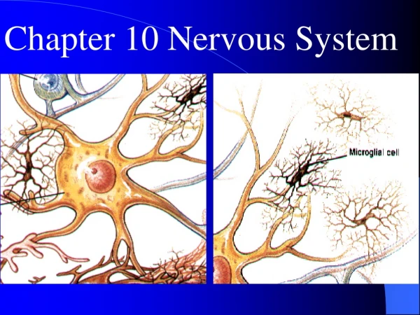
Neuroglia • Neuroglia or glial cells: • The nerve glue, holds the cell together. • Most glial cells are located in the CNS. • They support, insulate, nourish and care for delicate neurons. • Some participate in phagocytosis, others assist in the secretion of cerebrospinal fluid.
Neuroglia • Do not conduct electrical impulses. • Astrocytes- most abundant glial cell. • Supports the neurons and forms a protective barrier around the neurons of the CNS. • Barrier helps to prevent toxic substances in the blood stream from entering the nervous tissue of the brain and the spinal cord.
Neuron • Second type of nerve cell. • Most important in the transmission of information. • Enables the nervous system to act as a vast communication network. • Have many shapes and sizes. • Nonmitotic, do not replicate when injured.
Parts of a neuron • Parts of a neuron: • (1) Dendrites • (2) Cell Body • (3) Axon • (4) Axon Terminals
Parts of a Neuron • Axon-a long extension that transmits information away from the cell body. • The end of the axon undergoes extensive branching to form hundreds of thousands of axon terminals, this is where chemical neurotransmitters are stored.
Parts of a neuron • Myelin sheath-layer of white fatty material that encases most of the nerve fibers of the peripheral and central nervous system. • Myelin-protects and insulates the axon. • Myelinated-when nerve fibers are covered with myelin. • Unmyelinated-neurons that are not encased in myelin.
Types of Neurons • Three types of neurons: • (1) Sensory neuron-carries information from the periphery toward the CNS. Also called, afferent neurons. • (2) Motor neuron-carries information from the CNS toward the periphery. Also called efferent neurons. • (3) Interneuron-found only in the CNS. Form connections between sensory and motor neurons. In the brain, they play a role in thinking, learning and memory.
White matter and gray matter • The tissue of the CNS is white and gray. • White matter is white because of the myelin. • Myelinated fibers are gathered together in the CNS tracts. • The gray matter is composed primarily of cell bodies, interneurons, and unmyelinated fibers.
White matter versus gray matter • Nuclei-clusters of cell bodies located in the CNS. • Ganglia-(singular-ganglion)-small clusters of cell bodies located in the CNS. • Basal nuclei-patches of gray, located in the brain.
Nerve Impulses • Neurons allow the nervous system to rapidly convey information from one body part to the next. Ex. Stubbed toe. • Information is carried along the neuron in the form of a nerve impulse. • Nerve Impulse-an electrical signal that conveys information along a neuron.
Nerve Impulse • Action potential-a process of polarization, depolarization, and repolarization. • Polarization-the resting state of a neuron. No nerve impulse is being transmitted. The cell is quiet. • Depolarization-the neuron is stimulated, a change occurs in the cell’s electrical state. • Repolarization-cell returning to its resting place. Unless the cell repolarizes, it cannot be stimulated again. • Refractory Period-the cell’s unresponsive period. • The phases of the nerve impulse are caused by the movement of ions, particularly Na+ and K+.
Nerve impulse • Axons of most fibers are wrapped in myelin. • At the nodes of Ranvier, the axonal membrane is bare, not covered with myelin. • The nerve impulse arrives at the axon, it cannot develop on any part that is covered with myelin.
Nerve Impulse • To convey information, a nerve impulse must move the length of the neuron, from the cell body to the axon terminal.
Nerve Impulse • The nerve impulse will jump from node to node to the end of the axon. • Jumping from node to node is called saltatory conduction. • Saltatory conduction increases the speed with which the nerve impulse travels.
Events of a synapse • Many axons are myelinated to increase the speed of the nerve impulse. • The nerve impulse travels along the neuron from the dendrite to the end of the axon. • The impulse stimulates the release of neurotransmitters into the synaptic cleft. • The transmitter diffuses across the synaptic cleft, binds to the receptor and stimulates the dendrite of the second neuron.
The Brain • The brain is located in the cranial cavity. • Pinkish-gray, delicate structure with a soft consistency. • The surface of the brain appears bumpy, like a walnut. • Weighs about 3lb.
The Brain • Primary source of energy for the brain is glucose. • Low blood glucose levels result in hypoglycemia. • S/S patient will exhibit are: mental confusion, dizziness, convulsions, loss of consciousness, death.
The Brain • The brain is divided into four major areas: • The Cerebrum • The Diencephalon • The Brain Stem • The Cerebellum
The Cerebrum • The cerebrum contains both gray and white matter. • Cerebral Cortex-thin layer of gray matter that forms the outermost portion of the cerebrum. • The gray matter of the cerebral cortex allows us to perform higher mental tasks such as learning, reasoning, language, and memory. • The bulk of the cerebrum is composed of white matter located directly below the cortex.
The Cerebrum • The bumps of the cerebrum have several markings, or structures with special names. • The surface of the cerebrum is folded into elevations called convolutions or gyri.
The Cerebrum • The extensive folding increases the amount of the cerebral cortex. • It is thought that intelligence is related to the amount of cerebral cortex. • Sulci-the grooves that separate the gyri.
Lobes of the Cerebrum • Frontal Lobe- located in the front of the cranium under the frontal bone. • Plays a key role in voluntary motor function, personality, behavior, emotional expression, intellectual functions, and memory storage. Also involved with thinking learning and making plans. These are called “executive functions”
Frontal Lobe • Broca’s area-the part of the frontal lobe concerned with speech. • In most people it is in the left hemisphere. (some people it is in the right hemisphere). • If damaged, (CVA) the person develops aphasia – the person knows what they want to say but can’t
Frontal Lobe • Frontal eye field-located above Broca’s area. • Controls voluntary movements of the eyes and the eyelids. • Ex. Ability to scan a paragraph.
Decussation • Decussation-the crossing over of nerve fibers from one side of the brain to the other side of the body. • Fibers leave the motor area of the left frontal lobe cross over, and innervate the right side of the body. • The fibers from the right frontal lobe also cross over and innervate the left side of the body. • Ex. Damage to the left side of the brain causes paralysis to the right side of the body.
Parietal Lobe • Located behind the central sulcus. • Primarily concerned with receiving general sensory information from the body. • Called the primary somatosensory area because it receives sensations from the body.
Parietal Lobe • The primary somatosensory area- receives information from the skin and muscles and allows you to experience the sensations of temperature, pain, light touch, and proprioception, (a sense of where your body is). • The parietal lobe is also concerned with reading, speech and taste.
Temporal Lobe • Located inferior to the lateral fissure in the area above the ear. • Contains the primary auditory cortex, the area that allows you to hear. • Receives sensory information from the ears. • Damage to the temporal lobe causes deafness.
Temporal Lobe • Olfactory area-receives sensory information from the nose. Area that controls smell. • Sensory information from the taste buds- are located in the tongue. Interpreted in both the temporal and parietal lobes. • Wernicke’s area-broad region located in both parietal and temporal lobes; concerned with the translation of thoughts into words. • Damage to this area can cause deficits in language comprehension.
Occipital lobe • Located in the back of the head, underlying the occipital bone. • Contains the visual cortex. • Sensory fibers from the eye send information to the visual cortex of the occipital lobe, where it is interpreted as sight.
Occipital Lobe • Concerned with visual reflexes and vision-related functions such as reading. • Damage to the occipital lobe causes blindness.
Association areas • Large areas of the cerebral cortex that are concerned primarily with analyzing, interpreting and integrating information from the ear.
Patches of gray • Basal Nuclei-gray matter scattered throughout the cerebral white matter. • Help to regulate body movement and facial expression. • Dopamine is largely responsible for the activity of the basal nuclei. • A deficiency in dopamine within the basal nuclei is called Parkinson’s Disease.
Parkinson’s Disease • Characterized by: • A shuffling and uncoordinated gait. • Rigidity • Slowness of speech. • Drooling. • Masklike facial expression. • Usually treated with dopamine or dopamine-like drugs.
Diencephalon • Second main area of the brain. • Located beneath the cerebrum above the brain stem. • Includes the thalamus and the hypothalamus.
Thalamus • Serves as a relay station for most of the sensory fibers traveling from the lower brain and spinal cord region to the sensory areas of the cerebrum. • The thalamus sorts out the sensory information, gives us a hint of the sensation we are to experience, and then directs the information to the specific cerebral areas for more precise interpretation.
Hypothalamus • Second structure in the diencephalon. • Situated below the thalamus. • Regulates body temperature, water balance, and metabolism.