Download
1 / 12
120 likes | 375 Views
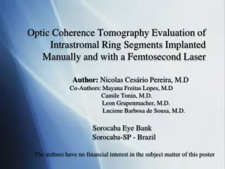
Optic Coherence Tomography Evaluation of Intrastromal Ring Segments Implanted Manually and with a Femtosecond Laser. Author: Nicolas Cesário Pereira, M.D Co-Authors: Mayana Freitas Lopes, M.D Camile Tonin, M.D. Leon Grupenmacher, M.D.

E N D
Optic Coherence Tomography Evaluation of Intrastromal Ring Segments Implanted Manually and with a Femtosecond Laser Author: Nicolas Cesário Pereira, M.D Co-Authors: Mayana Freitas Lopes, M.D Camile Tonin, M.D. Leon Grupenmacher, M.D. Luciene Barbosa de Sousa, M.D. Sorocaba Eye Bank Sorocaba-SP - Brazil The authors have no financial interest in the subject matter of this poster
Introduction • Intrastromal corneal ring segments have been implanted for 14 years with the manual tunnelization technique • Induces central cornea aplanation • Indications: Keratoconus, MPD, Post-LASIK ectasia, post PK , post RK, post trauma • Reversible, adjustable with fast visual reabilitation • Femtosecond Laser can be used for tunnelization • AS-OCT can be used to evaluate ring segments depth
Purpose • To evaluate intrastromal corneal ring segment depth with a high-speed corneal optical coherence tomography (OCT) system and to compare two techniques to implant the rings: the manual technique and assisted by the femtosecond laser
Setting • Hospital Oftalmologico De Sorocaba, Sorocaba Eye Bank, Sorocaba, Brazil. All the surgeries, exams and this study were carried out at Sorocaba Eye Bank.
Methods • Retrospective review of 34 patients submitted to intrastromal corneal ring implantation at Sorocaba Eye Bank between 2006 and 2008. Prospective evaluation with OCT was performed at least 3 months after the procedure. • Statistical analysis: t test, block variable analysis and Pearson correlation analysis
Methods • Measurements with OCT-Visante: • KeraringR: Triangular shape with base angle = 0o • Images with High Resolution Corneal Quad • Measurements realized with Caliper • 3 measurements: M1: Anterior cornea surface to apice of implant M2: Base of implant to posterior cornea surface M3: Full thickness cornea measurement central to the implant
Results • 41 eyes: 39 Keratoconus and 2 MPD • 24 implanted manually; 17 with FS • FS: Deeper and more regular • Planed depth was closer achived with FS • No statistical diference in clinical results • Complications: 3 extrusions (12,5%) with manual technique; no complications with FS • In 2 patients with FSOCT couldn’t measure residual cornea, but the patients were OK.
AV= acuidade visual; c/c=com correção; EE= equivalente esférico; Prof.=profundidade; ANP= anel nasal proximal; ANC=anel nasal central; AND= anel nasal distal; ATP= anel temporal proximal; ATC= anel temporal central; ATD= anel temporal distal.
MANUAL FS • No relation with VA and depth of implants
Results Manual Femtosecond Laser
Conclusions • OCT showed to be useful to evaluate the depth and regularity of intracorneal ring segments • No difference in the clinical results • Rings deeper and more regular with the FS • This can lead to less extrusion rate and less complications with FS.