Download
1 / 33
450 likes | 1.52k Views
Lecturer : Levkiv Mariana Department of Therapeutic Dentistry TSMU. Root canal filling materials. Root filling techniques. Purpose of root canal filling. To prevent bacteria and bacterial elements from spreading from (or through) the canal system to the periapical area ,

E N D
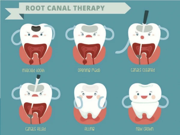
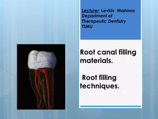
Lecturer: Levkiv Mariana Department of Therapeutic Dentistry TSMU Root canal filling materials. Root filling techniques.
Purpose of root canal filling • To prevent bacteria and bacterial elements from spreading from (or through) the canal system to the periapical area, • the fully instrumented root canal has to be provided with a tight and long-lasting obturation. • A root canal filling material should, therefore, prevent infection/reinfection of treated root canals. Together with an acceptable level of biocompatibility (inert material) this will provide the basis for promoting healing of the periodontal tissues and for maintaining healthy periapical conditions.
Instruments for root canal filling • Lentulo spiral filler/rotary paste filler • Function and features • • Small flexible instrument used to place materials into the canal • • Fits into the conventional handpiece • • Use with caution as it can be easily broken • • Different sizes available
Finger spreader Function, features and precaution • • Used to condense guttapercha into the canal during obturation • • Finger instrument with a smooth, pointed, tapered working end • • Disposed of in the sharps’ container Varieties • Can be of the hand instrument type (lateral condenser)
Endodontic plugger Function • Working end is flat to facilitate plugging or condensing the guttapercha after the excess • has been removed by melting off with a heated instrument Varieties • • Different sizes of working ends are available • •Available as hand or finger instruments
Guttapercha points Function and features • •Non-soluble, non-irritant points that are condensed into the pulp chamber during obturation • • Standardised type: follows same ISO classification as endodontic files • • Non-standardised: have a greater taper than the standard ISO type Varieties • • Can be packaged in single dose or bulk packages • • Different sizes with different tapers available
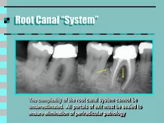
OBTURATING MATERIALSSealers • Regardless of the obturation technique employed, sealers are an essential component of the process. Sealers fill the space between the canal wall and core obturation material and may fill lateral and accessory canals, isthmuses, and irregularities in the root canal system.
The ideal properties of endodontic sealer are as follows: • 1. It should be tacky when mixed to provide good adhesion between it and the canal wall when set. • 2. It should produce a watertight seal • 3. It should be radiopaque so that it can be visualized o on X-ray. • 4. The particles of powder should be very fine so they can mix easily with the liquid. • 5. It should not shrink on setting. • 6. It should not stain tooth structure. • 7. It should be bacteriostatic or at least not encourage bacterial growth. • 8. It should set slowly. • 9. It should be insoluble in tissue fluids. • 10. It should be tissue-tolerant, that is nonirritating to periradicular tissue. • 11. It should be soluble in a common solvent in case removal of the root canal filling becomes necessary.
The most popular sealers are grouped by type: • Zinc oxide-eugenolformulations, • Calcium hydroxide sealers, • Glass- ionomers, and • Resins. Regardless of the sealer selected, all are toxic until they set. For this reason, extrusion of sealers into the periradicular tissues should be avoided.
Zinc oxide-eugenoland resin sealers have a history of successful use over an extended period. Zinc oxide-eugenolsealers have the advantage of being resorbed if extruded into the periradicular tissues . • Calcium hydroxide sealers were recently introduced for their potential therapeutic benefits. In theory these sealers exhibit an antimicrobial effect and have osteogenicpotential. Unfortunately these actions have not been demonstrated, and the solubility required for release of calcium hydroxide and sustained activity is a distinct disadvantage. • Glass ionomershave been advocated for use in sealing the radicular space because of their dentin bonding properties. A disadvantage is their difficult removal if retreatment is required.
Sealers containing paraformaldehyde are contraindicated in endodontic treatment. Although the lead and mercury components have been removed from the formulations over time, the paraformaldehyde content has remained constant and toxic. These sealers are not approved by the U. S. Food and Drug Administration.
Controversy surrounds removal of the smear layer before obturation. The smear layer is created on the canal walls by manipulation of the files during cleaning and shaping procedures. It is composed of inorganic and organic components that may contain bacteria and their by-products. In theory remnants left on the canal wall may serve as irritants or substrates for bacterial growth or interfere with the development of a seal during obturation. Although fluid movement may occur in obturated canals, bacterial movement does not appear to take place. Recent evidence suggests that removal of the smear layer can enhance penetration of the sealer into the dentinal tubules.
Removal of the smear layer can be accomplished after cleaning and shaping by irrigation with 17% ethylenediaminetetraacetic acid (EDTA) for 1 minute. Irrigation should be followed with a final rinse of sodium hypochlorite.
Acceptable methods of placing the sealer in the canal include the following: • • Placing the sealer on the master cone and pumping the cone up and down in the canal • • Placing the sealer on a file and spinning it counter clockwise • • Placing the sealer with a lentulo spiral • • Using a syringe • • Activating an ultrasonic instrument The clinician should use care when placing sealer in a canal with an open apex to avoid extrusion.
Core Obturation Materials • Historically, a variety of materials have been employed to obturate the root canal, falling into three broad categories: • solids, • semisolids, and • Pastes(sealers)
Sealers • A wide variety are available. The calcium hydroxide materials (e.g. Sealapex) or the eugenol-based sealers (e.g. Tubliseal) are perhaps the safest choice. Some would advocate the routine use of non-setting calcium hydroxide paste (Hypocal) as an inter-appointment medicament. • Calcium hydroxide This is considered separately, because it has a wide range of applications in endodontics due to its antibacterial properties and an ability to promote the formation of a calcific barrier. The former is thought to be due to a high pH and also to the absorption of carbon dioxide, upon which the metabolic activities of many root-canal pathogens depend. It is also proteolytic.
Indications for the use of calcium hydroxide include: • • To promote apical closure in immature teeth. • • In the management of perforations. • • In the treatment of resorption. • • As a temporary dressing for canals where filling has to be delayed. In the management of recurrent infections during RCT.
Solid materials • Silver cones met many of the criteria for filling materials but suffered from several deficiencies. The rigidity that made them easy to introduce into the canal also made them impossible to adapt to the inevitably irregular canal preparation, encouraging leakage. When leakage occurred and the points contacted tissue fluids, they corroded, further increasing leakage.
Semisolid material • Gutta-percha, a semisolid material, is the most widely used and accepted obturating material. Gutta-percha is a natural product that consists of the purified coagulated exudate of mazer wood trees (Isonandra percha) from the Malay archipelago or from South America. • Typical composition of gutta-percha cones.
Gutta-percha does not adhere to the canal walls, regardless of the filling technique applied, resulting in the potential for marked leakage. Therefore, it is generally recommended that gutta-percha (used cold or heated) is used together with a sealer. For an optimal seal the sealer layer should generally be as thin as possible.
Root filling techniques • Solid core techniques • Single cone – Simple – Quick – Good length control – Round standard preparation required • Lateral compaction – Good length control – Not one compact mass of gutta-percha – Time-consuming technique – Supposed risk of root fracture • Softened core techniques • Warm lateral compaction – Moderate length control – Time-consuming t chnique – Heat may damage periodontium • Warm vertical compaction – Poor length control – Sealer extrusion – Heat may damage periodontium • Injection-molded gutta-percha – Quick technique – Poor length control – Heat may damage periodontium • Thermomechanical compaction – Quick technique – Poor length control – Heat may damage periodontium – Instrument fracture risk • Core carrier – Quick technique – Sealer extrusion – Gutta-percha may be stripped off carrier in curvature – Difficult to remove for retreatment – In combination with posts, inconvenient technique • Chloroform–resin – Quick technique – Potential health hazard effects on dental personnel with long-term use
Root canal filling technique.Solid core technique • Single cone The single-cone technique consists of matching a cone to the prepared canal. For this technique a type of canal preparation is advocated so that the size of the cone and the shape of the preparation are closely matched. When a gutta-percha cone fits the apical portion of the canal snugly, it is cemented in place with a root canal sealer. Although the technique is simple, it has several disadvantagesand cannot be considered as one that seals canals completely. After preparation, root canals are seldom round throughout their length, except possibly for the apical 2 or 3 mm. Therefore, the single-cone technique, at best, only seals this portion.
ColdlateralcondensationThisis a commonlytaughtmethodofobturationandisthegoldstandardbywhichothersarejudged. Thetechniqueinvolvesplacement of a masterpointchosentofit theapicalsectionofthecanal. Obturationoftheremainderis achievedbycondensationof smalleraccessorypoints. The stepsinvolvedare:
1.Select a GP masterpointtocorrespondwiththemasterapicalfileinstrument. Thisshouldfittheapicalregionsnuglyattheworkinglengthsothatonremoval a degreeofresistanceor'tug-back'isfelt. Ifthereisno tug-backselect a largerpointorcut 1 mmat a timeoffthetipofthepointuntil a goodfitisobtained. Thepointshouldbenotchedatthecorrectworkinglengthtoguideitsplacementtotheapicalconstriction. • 2 .Take a radiographtoconfirmthatthepointisincorrectpositionifyouareinanydoubt. • 3.Coatwallsofcanalwithsealerusing a smallfile. • 4.Insertthemasterpoint, coveredincement. • 5 .Condensethe GP laterallywith a fingerspreadertoprovidespaceintowhichaccessorypointscanbeinserteduntilthecanalisfull. • 6.Excess GP iscutoffwith a hotinstrumentandtheremainderpackedverticallyintothecanalwith a coldplugger.
Sketch showing a cross-sectional cut through a root canal filled with a master cone and multiple accessory cones
Warm lateral condensation As above, but uses a warm spreader after the initial cold lateral condensation. Finger spreaders can be heated in a flame or a special electronically heated device (Touch of heat) can be used.
Verticalcondensation • In this technique the GP is warmed using a heated instrument and then packed vertically. A good apical stop is necessary to prevent apical extrusion of the filling, but with practice a very dense root filling can result. Time consuming.
Diagram of the warm vertical condensation technique. • A, After a heated spreader is used to remove the coronal segment of the master cone, a cold plugger is used to apply vertical pressure to the softened master cone. • B,Obturation of the coronal portion of the canal is accomplished by adding a gutta -percha segment. • C, A heated spreader is used to soften the material. • D, A cold plugger is then used to apply pressure to the softened gutta-percha.
ThermomechanicalcompactionThisinvolves a reverseturning (e.g. McSpaddencompactoror GP condenser) instrumentwhich, like a reverseHedstroemfile, softensthe GP, forcingitaheadof, andlateraltothecompactorshaft. Thisis a veryeffectivetechnique, particularlyifusedinconjunctionwithlateralcondensationintheapicalregion, butrequiresmuchpracticetoperfect. • Thermoplasticizedinjectable GP (e.g. Obtura, Ultrafil)Thesecommercialmachinesextrudeheated GP (70-160°C) intothecanal. Itisdifficulttocontroltheapicalextentoftherootfilling, andsomecontractionofthe GP occursoncooling. Usefulforirregularcanaldefects, e.g. followinginternalrootresorption.
Coatedcarriers (e.g. Thermafil) Thesearecoresofmetalorplasticcoatedwith GP. Theyareheatedinanovenandthensimplypushedintotherootcanaltothecorrectlength. Thecoreisthenseveredwith a bur. A densefillingresults, butagainapicalcontrolispoorandextrusionscommon. Theyareexpensiveanddifficulttoremove. • Oncethefillingisinplacethetoothwillneedtobepermanentlyrestored, providedthe follow-up radiographissatisfactory. Fillingsthatappearinadequateradiographicallymaybereviewedregularly, orreplaced, dependingupontheclinicalcircumstances.
THE CORONAL SEAL • Regardless of the technique used to obturate the canals, coronal microleakage can occur through seemingly well- obturated canals within a short time, potentially causing infection of the periapical area. A method to protect the canals in case of failure of the coronal restoration is to cover the floor of the pulp chamber with a lining of glass ionomer cement after the excess gutta-percha and sealer have been cleaned from the canal. Glass ionomers have the intrinsic ability to bond to the dentin, so they do not require a pretreatment step. The resin-modified glass ionomer cement is simply flowed approximately 1 mm thick over the floor of the pulp chamber and polymerized with a curing light for 30 seconds. Investigators found that this procedure resulted in none of the experimental canals showing leakage