Download
1 / 10
100 likes | 235 Views
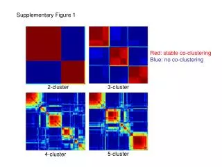
Supplementary Figure 1. Red: stable co-clustering Blue: no co-clustering. 2-cluster. 3-cluster. 5-cluster. 4-cluster. Supplementary Figure 2. Supplementary Figure 3. EGFR mutation. P=0.16. Supplementary Figure 4. Supplementary Figure 5. EGFR mutation. Supplementary Figure 6.

E N D
Supplementary Figure 1 Red: stable co-clusteringBlue: no co-clustering 2-cluster 3-cluster 5-cluster 4-cluster
EGFR mutation P=0.16 Supplementary Figure 4
EGFR mutation Supplementary Figure 6 KRAS mutation
Supplementary Figure 7 p=0.0001 DUSP4 deletion p16 deletion 26 10 16 14 5 7 p=0.00003 p=0.007 17 104 EGFR mutation n=199
Supplementary Figure 8 6E-16 Wild type EGFR EGFR L858R EGFR del19 5E-16 4E-16 DUSP4 mRNA conc (uM) 3E-16 2E-16 1E-16 0 WT - 2 hrs WT - 4 hrs WT - No EGF WT - 6 hrs WT - 1 hr WT - 30 min del19 - No EGF del19 - 2 hrs del19 - 6 hrs del19 - 4 hrs L858R - No EGF del19 - 30 min L858R - 2 hrs L858R - 4 hrs del19 - 1 hr L858R - 30 min L858R - 6 hrs L858R - 1 hr
Supplementary Figure 9 A B DUSP4-GFP DUSP4 GFP Vector 70KD DUSP4-GFP GFP 27KD ACTIN H358 H1650 H1650
Supplementary Figure 10 H1650 cells 24hrs after electroporation H1650 cells after one week selection GFP DUSP4-GFP