Download
1 / 14
140 likes | 198 Views
This text provides an overview of different methods and assays used for protein isolation, purification, and identification. It covers topics such as protein solubilization, sub-cellular fractionation, protein assays, and purification strategies.

E N D
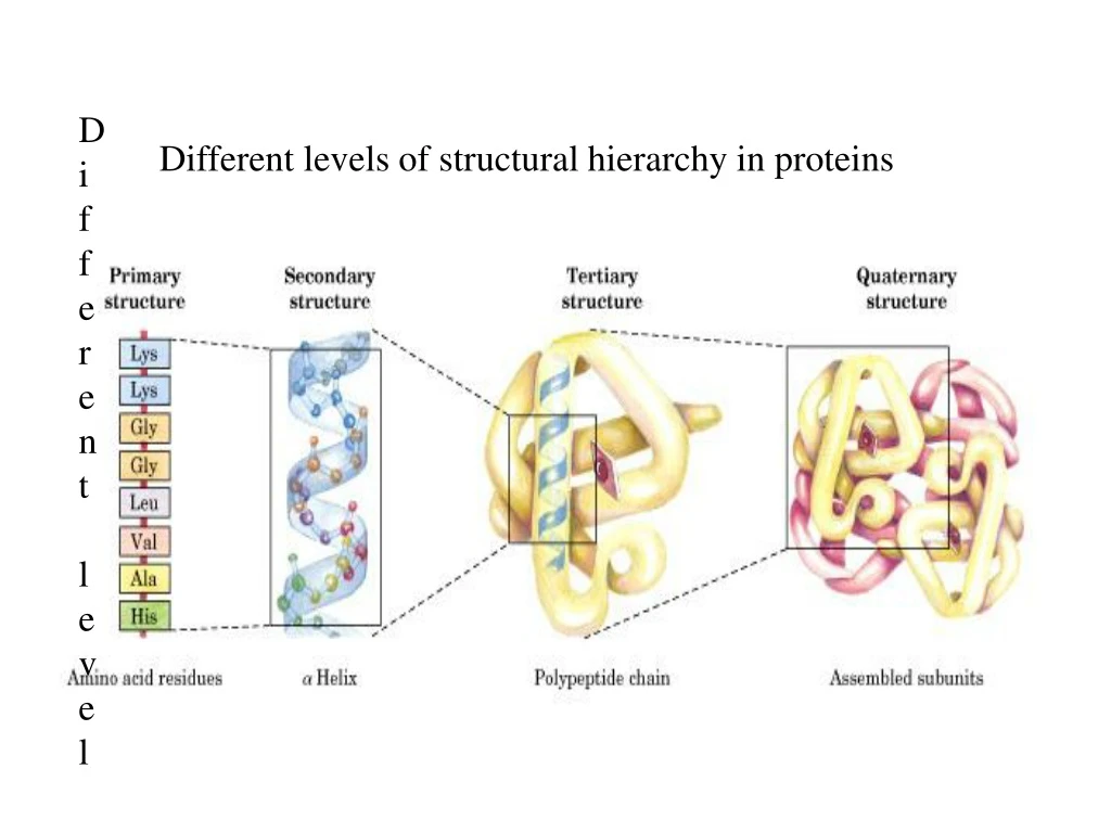
Different level of Different levels of structural hierarchy in proteins
Working with Proteins: • Protein Isolation, Purification and Identification • Protein source: Examples: Tissue samples, plant materials, cultured mammalian or plant cells yeast, bacteria • Solubiliozation of Proteins: • Homogenization of the source material: • Cell rupture by hypotonic solution followed by homogenization • Cell lysis by detergents followed by homogenization • For yeast and bacteria rigorous grindings and enzymatic treatments • are used to break the cells.
Sub-cellular fractionation: To obtain and study proteins from various • cellular compartments, cells are ruptured by hypotonic shocks to keep • all membranes intact. A differential centrifugation protocol is used to • Separate fractions e.g. nuclear, mitochondrial, membranous and • fraction. See next figure. • Once proteins are extracted out from their cellular environments • They are unstable: • May get denatured (loss of structure and activity) • May get proteolysed by several proteases released from cell • rupture • May get modified (oxidized) • To avoid this most of the time, all prification steps are carried out • At 0-4 oC and in presence of general protease inhibitors.
Assay of proteins: • Total proteins and • specific protein which is under investigation (by enzymatic activity or specific probe) • Total protein amount can be assayed by • Biuret assay • Lowry Method • Both these methods are based on the binding of Cu2+ with the N of peptide bond in alkaline condition producing tetra-dentate complex • This complex produces blue colour which can be measured (absorption at 550 for biuret and 750 nm for lowry’s method). Increase in absorption is proportional to the anount of proteion. • Coomassie Blue Dye binding assay (Bradford Method): This dye binds to protein in acidic condition.
There should be some way of assessing the specific protein being Purified during each step of purification. For proteins with enzymatic activities such as peroxidase, phosphatase’ Protease, nucleases, polymerases etc, there are convenient assays These assays use substrates which are converted to fluorogenic product or product with different absorption maxima (colourimetric product). Measurement of the amount of product in reaction mixture is correlated with the amount of protein enzyme. For proteins without any enzymatic activity, specific probes like antibodies to them are used to quantify the amount of protein. ELISA is one of the most common procedure used in diagnostic and Research laboratories to quantify specific proteins or peptides.
Radioimmume assy (RIA): It is very sensitive technique to estimate the Amount of known protein/peptide in any solution. Using a known concentration of radiolabelled peptide and immobilized antibody, the amount of the unlabelled peptide in unknownsolution is estimated. The unlabelled peptide in the sample competes with labelled one. Amount of radioactive peptide displaced (which can be measured easily) by unlabelled peptide (the unknown concentration) corresponds to the amount of unlabelled peptide. A standard curve using different known amount of unlabelled peptide in this assay can be made. By putting the value of radioactive peptide released by unknown solution on standard curve one can easily calculate concentration of peptide in sample solution.
Strategy for purification of a protein: • Once the protein extract is prepared, in order to prify a single protein • from this extract different fractionation methodologies can be used. • Salting in and salting out • pH dependent precipitation • Protein charge based methods: Ion exchange chromatography, • electrophoresis, and isoelectric focussing • Protein size based methods: Dialysis, ultrafiltration, gel • Electrophoresis, gel filtration chromatography and ultrafiltration • Polarity based methods: Reverse phase chromatography, • Hydrophobic interaction chromatography, adsorption • chromatography
Effect of salt concentration on the solubility of proteins Ionic strength (I) of salt affects the solubility of proteins. I = 1/2 ciZi2 Ci is molar concentration of the ionic species Zi is the charge of the ion Salting in: As the salt concentration (ionic strength) increases, the Additional ions more effectively shield the protein molecule’s multiple charges increasing its solubility. Different proteins get solubilized at different ionic strengths. (see figure 5-2 of the book)
Salting out: At higher ionic strength, the solubility of proteins decreases. This because, the availability of solvent molecules becomes limited as Most of them are used up by salt ions. Ammonium sulphate is the most preferred salt used in biochemistry For salting out proteins. This is because it has very high solubility (close To 4 M at zero degree Celsius). Refer to fig. 5-3 in the book. Effect of pH on solubility: In solution of moderate salt solution, the solubility proteins are minimum at pH around the isoelectric Point (pI) of the protein. Some proteins are fractionated using this chriterion