Download
1 / 38
380 likes | 413 Views
Explore a neurology case study to identify the origin of symptoms through neuraxis divisions and neurologic examinations. Understand lesions in the cortex, subcortex, brainstem, and cerebellum. Learn to assess cortical brain functions and subcortical brain abnormalities.

E N D
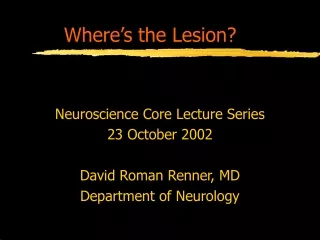
Where’s the Lesion? Neuroscience Core Lecture Series 23 October 2002 David Roman Renner, MD Department of Neurology
Scott’s CC: • “My balance is off.” • Multiple ER visits for fall-related trauma • “I’m losing the fine control of my fingers.” • Loss of manual dexterity • “I’ve had pneumonia three times.” • Dysphagia to liquids>solids
All of Scott’s Complaints Sounded Neurologic in Origin His lesion should lie somewhere in the neuraxis.
Divisions of the Neuraxis • Cortical Brain • Subcortical Brain • Brainstem • Cerebellum • Spinal Cord • Root • Peripheral Nerve • Neuromuscular Junction • Muscle
Off the Top of my Head . . . • Imbalance = Cerebellum • Pneumonia = Brainstem (related dysphagia) • Loss of Dexterity = Peripheral Nerve
Neurologic Examination • Higher Cortical Function • Cranial Nerves • Cerebellar Function • Motor • Sensory • Deep Tendon Reflexes • Pathologic Reflexes
The Neuro Exam Should Evaluate the Entire Neuraxis • Higher Cortical Function: cortex • Cranial Nerves: subcortex, brainstem • Cerebellar Function: cerebellum • Motor: motor homonculous, subcortical pyramidal tracts, BS, cord, radicle, PN, muscle • Sensory: ascending tracts, thalamus, subcortical tracts, sensory hononculous • Deep Tendon Reflexes: afferent PN, radicle, cord, efferent PN, muscle • Pathologic Reflexes:
Scott’s Exam Showed: • Higher Cortical Function: normal • Cranial Nerves: oropharyngeal dysarthria • Cerebellar Function: hypotonia, assynergy, dysmetria, staccato dysarthria, intention tremor, appendicular ataxia • Motor: hypotonia, normal strength • Sensory: decreased vibration and temperature • Deep Tendon Reflexes: areflexia • Pathologic Reflexes: plantar flexing
Divisions of the Neuraxis • Cortical Brain • Subcortical Brain • Brainstem • Cerebellum • Spinal Cord • Root • Peripheral Nerve • Neuromuscular Junction • Muscle
Cortical Brain • Depends upon hemispheric dominance • Non-neurologists generalize: • right: visual/spatial, perception and memory • left: language and language dependent memory • Look for aphasias, apraxias, and agnosias
Neurologic Examination when Cortical Brain is Lesioned • Higher Cortical Function: aphasia, apraxia, agnosia • Cranial Nerves: normal • Cerebellar Function: normal • Motor: weakness if you hit the motor homonculous • Sensory: sensory abnormalities if you hit the sensory homonculous • Deep Tendon Reflexes: hemi-hyper-reflexia • Pathologic Reflexes: possibly Babinski’s reflex or frontal release signs
Divisions of the Neuraxis • Cortical Brain • Subcortical Brain • Brainstem • Cerebellum • Spinal Cord • Root • Peripheral Nerve • Neuromuscular Junction • Muscle
Subcortical Brain • Deep white radiating fibers produce equal involvement of face/arm/leg • weakness • sensory abnormalities • Visual radiating fibers are interrupted: • deep parietal: pie on the floor • deep temporal: pie in the sky
Neurologic Examination when Subcortical Brain is Lesioned • Higher Cortical Function: normal • Cranial Nerves: visual field cuts • Cerebellar Function: usually normal • Motor: weakness in face=arm=leg, UMN • Sensory: sensory abnormalities in face=arm=leg • Deep Tendon Reflexes: hemi-hyper-reflexia • Pathologic Reflexes: Babinski’s reflex and possibly frontal release signs
Divisions of the Neuraxis • Cortical Brain • Subcortical Brain • Brainstem • Cerebellum • Spinal Cord • Root • Peripheral Nerve • Neuromuscular Junction • Muscle
Brainstem • The Brainstem is basically spinal cord with embedded cranial nerves, producing the following abnormalities • cranial nerve abnormalities • classic spinal cord complaints • bowel and bladder problems • long tract signs: (bilateral and crossed) • corticospinal (pyramidal): motor • spinothalamic: pain/temp to the thalamus • dorsal columns: prioprioception/vibration to thal.
Neurologic Examination when Brainstem is Lesioned • Higher Cortical Function: normal • Cranial Nerves: • III, IV, VI: diplopia • V: decreased facial sensation • VII: drooping • VIII: deaf and dizzy • IX, X, XII: dysarthria and dysphagia • XI: decreased strength in neck and shoulders • Cerebellar Function: normal • Motor: hemi-paresis, UMN • Sensory: hemi-dysesthesias • Deep Tendon Reflexes: hemi-hyper-reflexia • Pathologic Reflexes: Babinski’s reflex
Divisions of the Neuraxis • Cortical Brain • Subcortical Brain • Brainstem • Cerebellum • Spinal Cord • Root • Peripheral Nerve • Neuromuscular Junction • Muscle
Cerebellar Function • Some people believe that one can not test specifically for cerebellar abnormalities • no one test on examination reliably evaluates the cerebellum • H: hypotonia • A: assynergy of (ant)agonist muscles • N: nystagmus • D: dysmetria, dysarthria • S: stance and gait • T: tremor
Neurologic Examination when the Cerebellum is Lesioned • Higher Cortical Function: normal • Cranial Nerves: normal • Cerebellar Function: • nystagmus • staccato dysarthria (abnormality of prosody) • Motor: • hemi-hypotonia • intention > positional tremor • axial instability with dysmetria • Sensory: normal • Deep Tendon Reflexes: normal • Pathologic Reflexes: none
Divisions of the Neuraxis • Cortical Brain • Subcortical Brain • Brainstem • Cerebellum • Spinal Cord • Root • Peripheral Nerve • Neuromuscular Junction • Muscle
Spinal Cord • Sensory level (horizontal) • Weakness below the lesion (paraparesis) • UMN signs below the lesion • Bowel and bladder incontinence
Neurologic Examination when the Spinal Cord is Lesioned • Higher Cortical Function: normal • Cranial Nerves: normal • Cerebellar Function: normal • Motor: weakness below the lesion • Sensory: horizontal level • Deep Tendon Reflexes: hyper-reflexia below the lesion • Pathologic Reflexes: Babinski’s reflex
Divisions of the Neuraxis • Cortical Brain • Subcortical Brain • Brainstem • Cerebellum • Spinal Cord • Root • Peripheral Nerve • Neuromuscular Junction • Muscle
Root/Radiculopathy • Painis the hallmark of a radiculopathy • Sensory abnormalitiesin a dermatome • provocative maneuvres exacerbate the pain • Weaknessin a myotome (assymetric) • LMN findings
Neurologic Examination when a Root is Lesioned • Higher Cortical Function: normal • Cranial Nerves: normal • Cerebellar Function: normal • Motor: assymetric weakness in a myotome • Sensory: pain and dysesthesia confined to a dermatome • Deep Tendon Reflexes: hypo- to a-reflexia if the root carries a reflex • Pathologic Reflexes: none
Divisions of the Neuraxis • Cortical Brain • Subcortical Brain • Brainstem • Cerebellum • Spinal Cord • Root • Peripheral Nerve • Neuromuscular Junction • Muscle
Peripheral Nerve(presuming nonfocality) • Weakness: distal predominant • Sensory Dysesthesias: distal predominant
Neurologic Examination with Diffuse PN Lesioning • Higher Cortical Function: normal • Cranial Nerves: normal • Cerebellar Function: normal • Motor: weakness is distal predominant • Sensory: dysesthesias are distal predominant • Deep Tendon Reflexes: loss of distal reflexes • Pathologic Reflexes: mute responses to plantar stimulation
Divisions of the Neuraxis • Cortical Brain • Subcortical Brain • Brainstem • Cerebellum • Spinal Cord • Root • Peripheral Nerve • Neuromuscular Junction • Muscle
Neuromuscular Junction • Fatiguabilityis the hallmark • Weakness: proximal and symmetric • exacerbated with use, recovers with rest • often affects facial muscles (ptosis, dysconjugate gaze, slack jaw) • Sensation: preserved
Neurologic Examination in Disorders of the NMJ • Higher Cortical Function: normal • Cranial Nerves: fatiguabile ptosis, dysconjugate gaze, slack jaw • Cerebellar Function: normal • Motor: fatiguable proximal weakness in both UE’s and LE’s • Sensory: normal • Deep Tendon Reflexes: normal • Pathologic Reflexes: none
Divisions of the Neuraxis • Cortical Brain • Subcortical Brain • Brainstem • Cerebellum • Spinal Cord • Root • Peripheral Nerve • Neuromuscular Junction • Muscle
Muscle • Weakness of proximal arm and leg muscles • symmetric • Sensation is normal • though patients complain of cramping and aching
Neurologic Examination in Disorders of Muscle • Higher Cortical Function: normal • Cranial Nerves: ptosis, dysconjugate gaze, dysphagia, dysphonia, (dysarthria) • Cerebellar Function: normal • Motor: proximal weakness in both UE’s and LE’s, atrophy and fasiculations, hypotonia • Sensory: normal • Deep Tendon Reflexes: preserved until late in the disease • Pathologic Reflexes: none
Scott’s Lesion Localizes to: Almost exclusively the Cerebellum, though to a minor degree the BS and PN are involved. Isolated heritable cerebellar dysfunction is rare, so we would expect to see other parts of the CNS involved.
SpinoCerebellar Ataxia (SCA4) Prior to Scott’s diagnosis, his cousin was the proband for this entity. • Scott has a 38-family member, 5 generation pedigree of this disorder His family entered into a study, and his family led to the classification of SCA4: ataxia with axonal sensory neuropathy