Download
1 / 43
440 likes | 698 Views
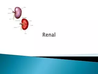
RNSG 2120 - Renal. Gwyn Brandi, MSN, RN, CMSRN Fall 2012. Kidneys. Functions Urine formation Excretion of waste Regulation of electrolytes Regulation of acid-base Water balance Control of BP Regulates red blood cell production Synthesis of Vit D. Review Anatomy

E N D
RNSG 2120 - Renal Gwyn Brandi, MSN, RN, CMSRN Fall 2012
Kidneys • Functions • Urine formation • Excretion of waste • Regulation of electrolytes • Regulation of acid-base • Water balance • Control of BP • Regulates red blood cell production • Synthesis of Vit D • Review • Anatomy • Blood supply to kidneys • Nephrons • Urine formation • Glomerular filtration • Functions of kidneys
GFR • GLOMERULAR FILTRATION RATE = KIDNEY FUNCTION • Glomerular filtration rate (GFR) is the volume of fluid filtered from the renal (kidney) glomerular capillaries into the Bowman‘s capsule per unit time. Normal GFR is 80-150 ml/hr
Renin-Angiotensin System • Normal – body senses the blood pressure and adjusts accordingly • Abnormal – body over-releases angiotensin and aldosterone
Urinalysis & U/A C&S • Urine Exam Includes • Urine color • Urine clarity & odor • Urine pH • Specific gravity • Tests for protein, glucose, ketones • Detect RBC’s, WBC’s, casts, crystals, & bacteria • Culture & Sensitivity • Usually takes 3 days • Allows bacteria to grow • Then growth is tested using a variety of ATBs • The report is then returned showing which ATBs work & which ones don’t
Specific Gravity • Normal range – 1.010 to 1.025 • Measures density of urine • Decrease = diabetes insipidus, glomerulonephritis, severe renal damage • Increase = diabetes mellitus, nephritis, fluid volume deficit (dehydration) • Fluid volume decreases or dehydration = increase in specific gravity (concentrated) • High fluid intake = decrease in specific gravity (diluted) • Tested by dipstick (quick, on floor, by nurse), or refractometer (used in the lab)
Urine Osmolality • Concentrating & diluting ability of the kidneys • Lost early in kidney disease • Test findings can determine deficits in kidney function/renal failure
24 Hour Urine • Determines progression of renal disease • You should collect every drop of urine during each 24-hour period • Urinate (empty the bladder) for the first time and flush it down the toilet. Note the exact time (eg, 6:15 AM). You will begin the urine collection at this time • Collect every drop of urine during the day and night in an empty collection bottle. Store the bottle at room temperature or in the refrigerator • Note the exact time of the final collection, even if it is not the same time as when collection began on day one
BUN & Creatinine • Blood Urea Nitrogen • Normal range – 7 to 18 • Serves as index of renal function • Determines ability of kidneys to process proteins • High levels indicate kidney function is less than normal • Creatinine • Normal range – 1.6 to 1.2 • Measures effectiveness of renal function • Should remain fairly constant in body • High levels indicate kidney damage
Nuclear Scan • Injection of radioisotope dye • Uses a camera placed behind kidney • Provides info on kidney perfusion (blood flow) & renal failure
Intravenous Pyelography • IV radiopaque dye • Under xray, shows dye moving through the upper & lower urinary system (kidneys, ureters, bladder)
Retrograde Pyelography • Catheters placed in ureters using cystoscope • Dye injected • Used if IV pyelography is inadequate or prior to extracorporeal shock wave lithotripsy
Renal Angiography • Also known as a Renal Arteriogram • Assesses the renal arteries • Catheter inserted in femoral artery and floated to the renal artery • Contrast dye is injected • Under angiography, the renal blood flow is evaluated • Used to evaluate renal trauma, renal cysts vs. tumors, pre-renal transplant, and renal hypertension • Watch for bleeding at site, peripheral pulses, hematoma formation, impaired renal function
Renal Biopsy • Brush Biopsy • Cystoscopy performed • Brush inserted and brushed against lesion or suspected tumor • Cells retrieved and tested • Kidney Biopsy • Used to assess renal failure, proteinuria, hematuria, transplant rejection, glomerulopathies • Needle biopsy or open biopsy • Local or general anesthetic • Inserted into the renal capsule
Nephrotic Syndrome • Type of renal failure characterized by increased glomerular permeability and is manifested by massive proteinuria • S/sx – increased albumin in urine, proteinuria, decrease albumin in blood, diffuse edema, high cholesterol, hyperlipidemia (LDL) • Overtaxes the kidneys, so the liver tries to compensate, but it can only do so much • Main s/sx is edema – pitting periorbital edema, dependent edema, peripheral edema, and ascites • Tx – diuretics, and ACE inhibitors to reduce proteinuria and LDL
Polycystic Kidney • Genetic disorder • Growth of numerous fluid filled cysts in the kidneys • Destroys the nephrons • Leads to renal failure • Can lead to cysts formation in liver, blood vessels, brain, and heart • S/sx – increased size of kidneys, hematuria, polyuria, hypertension, renal calculi, UTIs, proteinuria, back/flank pain
Polycystic Kidney • Dx – palpation, genetic testing, ultrasound • Tx – no cure, supportive therapy for symptoms, when kidneys fail - dialysis
Acute Renal Failure (ARF) • Rapid decline in renal function • S/sx – asymptomatic, detected by labs, oliguria, elevated BUN & creatinine • Prerenal cause – decreased bloodflow to kidney • Intrarenal cause – within the kidney • Postrenal cause – obstruction to urine flow • Near complete cessation of kidney function
Acute Renal Failure • Can lead to metabolic acidosis • Surgery • Diet • Low protein • Low phosphate • Avoid foods high in K+ • Tx • Treat underlying cause • Fluid replacement/restriction • Dialysis • Diuretics • I’s and O’s • Daily weights • Monitor labs, BP, & respiratory status
Medical Management - Prerenal • Early recognition • Fluid or volume replacement • Caution in patients with underlying cardiac disease • May require inotropes, antidysrrhythmic agents, preload/afterload reducers, intraaortic balloon pump • May require hemodynamic monitoring to guide treatment
Medical Management - Postrenal • Alleviate obstruction • May need stent • Dietary or lifestyle changes may be required
Medical Management - Intrarenal • Efforts aimed at prevention • Identify risk • Drug Therapy • Diuretics • Dopamine – may increase renal blood flow • Acetylcysteine • Theophylline • Epoetinalfa – to treat anemia
Chronic Renal Failure • ESRD – end-stage renal disease • Causes – DM, ARF, HTN • DX • Decreasing GFR • Creatinine clearance to determine GFR • CBC • Urinalysis • S/sx • Nausea • Anorexia • Dry mouth with uremic fetor • Clonic jerks • Pulmonary edema & acidosis • Tx • Dialysis • Renal transplant
Chronic Renal Failure • End stage • Uremic encephalopathy/coma • Immunosuppression • Volume overload • Metabolic acidosis • Hyperkalemia • Respiratory failure
Chronic Renal Failure • Pharmacology • Narcotics (morphine) • Phosphate binders (calcium carbonate) • Benzodiazepines for clonus • Epogen (Procrit) for anemia • Vitamin D supplements • Water soluble vitamin supplements • Schedule meds AFTER dialysis
Dialysis Dialysis does the following: • removes waste, salt and extra water to prevent them from building up in the body • keeps a safe level of certain chemicals in your blood, such as potassium, sodium and bicarbonate • helps to control blood pressure Dialysis Indications • Fluid overload • Electrolyte imbalances • Acid-base disturbances
Hemodialysis • AV shunt • Lower arm most common • Anastomosis • No BP’s or sticks • Bruit and thrill • Blood pumped through dialysate fluid and put back in circulation • 3 – 4 times a week, 3 – 4 hrs per session
Hemodialysis • Common complications • Steal syndrome • Infection • Loss of shunt • Steal syndrome – disruption of blood to extremity • Pain • Coolness • paresthesias
Vascular Access No blood pressure check No IV sticks Loose clothing
Peritoneal Dialysis • Dialysate fluid instilled in peritoneal cavity • Osmotic pressure to draw out waste materials • Instill fluid for a prescribed amount of time
Peritoneal Dialysis • Sterile technique with masks • Risk of peritonitis • Cloudy effluent (waste draining) • Abdominal pain • Rigid abdomen
Dialysis Dietary Managment Higher than normal basal requirement Provide adequate energy, protein, and micronutrient 25 to 35 kcal/kg of ideal body wt per day. Water soluble vitamins lost in dialysis • A Healthy Diet for a dialysis patient is: • adequate in protein • adequate in calories • low to moderate in potassium • low in sodium • low in phosphorus • controlled in fluids
Renal Transplant • Live or cadaver donor • Genetic testing – histocompatibility • Surface antigens (HLA), T-cells • Following transplant, watch urine output • Watch electrolyte balance • Immunosuppressants • Expensive
Diuretics • More fluid in the blood – the higher the blood pressure • Fluid goes out, blood pressure goes down
Loop Diuretics • I & O • Inhibits sodium & chloride in the loop of Henle • May cause dig toxicity when given concurrently • Lasix (furosemide) • Hypokalemia • Hypotension • Monitor K+ level
Thiazide Diuretics • Decrease reabsorption of sodium, water, chloride, & bicarb in the distal convoluted tubule • Not that strong of a diuretic • HCTZ • Useful in tx of HTN • Hypokalemia • Orthostatic hypotension • Monitor K+ level
Potassium Sparing Diuretics • Aldactone (spironolactone) • Hyperkalemia • Monitor Lithium levels • Give after meals • Weak diuretic – usually used in combination with other diuretics