Download
1 / 24
250 likes | 585 Views
Technology and The Future of Medicine. "Altering tumor cell scaffolding: Looking for cytoskeleton-damaging agents". Consolato Maria Sergi, MSc, MD, PhD Department of Laboratory Medicine and Pathology University of Alberta. 1. Outlines .

E N D
Technology and The Future of Medicine "Altering tumor cell scaffolding: Looking for cytoskeleton-damaging agents" Consolato Maria Sergi, MSc, MD, PhDDepartment of Laboratory Medicine and Pathology University of Alberta 1
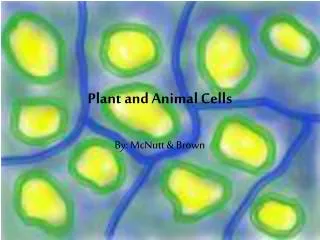
Outlines • Eukaryotic Cells contain many organelles and a complex cytoskeleton. • Cytoskeleton as scaffolding of a cell. • Actin cytoskeleton is organized into bundles and networks of filaments. • Intermediate filaments are dynamic polymeric in the cell. • Role of signal-transduction pathways in cell locomotion and the organization of the cytoskeleton. • Intermediate filaments in disease. • Possible anti-cancer therapy.
Microtubules • Located in the cytoplasm of all eukaryotic cells • 25 nm in diameter ( the thickest fiber type) • Built up of Tubulin (β-tubulin & α-tubulin) • Movements tracks • Separation of the chromosomes • Compression resistance 4
Microtubules: Centrosomes & Centrrioles • Where the microtubules are initiated 5
Microtubules: Cell and cell’s organelles movements • Cilia & Flagella:• protrude from cell surface • cilia: abundant number with 0.25μm in length• flagella: usually 1 – few per cell with 2-20 μmin length • “9+2” patterns 6
Microtubules: Cell and cell’s organelles movements • Beating patterns in cilia and flagella 7
Microtubules: Cell and cell’s organelles movements • Motor proteins: when the motor protein attaches to organelle, it has the ability to move the organelle along microtubule. 8
Microfilaments (Actin filaments) • Built from actin molecules • Mechanical support • 3D Structural networks insideplasma membrane • Cell motility: contractility of muscle cells • Actin & Myosin: the contractile engine 9
Actin & Myosin: the contractile engine • Myosin : Motor protein 10
Actin filaments • Actin cytoskeleton is organized into bundles and networks of filaments • Plasma membrane support 11
Actin filaments • Bundles: actin filaments are closely packed in parallel arrays • Networks: actin filaments are crisscross and loosely packed. 3D structure = gel-like cytosol • The filaments of the bundles and networks are connected by actin-cross-linking proteins 12
Actin filaments: actin-cross-linking proteins • The formation of the bundles or networks are highly depended on the flexibility of the cross-linking protein • Actin bundles: e.g. fimbrin & α-actinin • Actin networks: e.g. filamin & spectrin 13
Intermediate filaments (Ifs) • Diameter: microtubules>intermediat filament> microfilaments • More permanent fixtures of cells • Keratins • Don not have a role in cell motility 14
3D reconstruction of a cytoskeleton • It is highly useful tool to fully understand the structural and mechanical properties of the cytoskeleton Extraction of the graph structure from a detailed section of a secondary electron tomogram. (a) Detail of a tomogram after thresholding. (b) Skeleton of the foreground phase. (c) Extracted network graph after all processing steps, cross-links highlighted by spheres. 16 Journal of Microscopy, Vol. 239, Pt 1 2010, pp. 1–16
IF network in a detergent-extracted Panc 1 cell visualized at a magnification of 50,000. Filaments in some depth are clearly visible at good contrast in the secondary electron tomogram. The graph extracted from the tomogram exhibits a genuinely 3D structure. (a) SEM image at 0◦ tilt. (b) Horizontal cut through an SEM tomogram. (c) Vertical cut through an SEM tomogram. (d) Tomogram after binarisation (top view). (e) Network graph extracted from the tomogram, top view (left), rotated by 40◦ around central axis (right). 17 Journal of Microscopy, Vol. 239, Pt 1 2010, pp. 1–16
3D reconstruction of a cytoskeleton electron microscope stereo images (400]400 in pixels) of cell cytoskeleton taken at$103 of tilt angles Reconstructed 3D cytoskeleton structure visualized by isosurface method. 18 Journal of Biomechanics 33 (2000) 105}113
Role of signal-transduction pathways in cell locomotion and the organization of the cytoskeleton 19
Cont’d 21
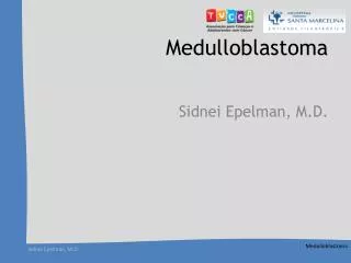
Hedgehog (Hh) signaling in cancer • Examples: Basal cell carcinomas Medulloblastomas 22 http://www.med.umich.edu/derm/research/res_embryonic.shtml
Inhibition of Hh-Gli-1signaling in cancers • Inhibition of human prostate cancer cell proliferation by cyclopamine and GLI1 RNA interference. Cyclopamine inhibits the proliferation of primary in situ prostate tumors (A) and of metastatic prostate cancer cell lines (C), as measured by BrdU incorporation, and inhibits expression of GLI1 (B). Similarly, GLI1 small interfering RNAs (siRNAs) decrease GLI1 and PTCH1 expression (D,F) and inhibit the proliferation of prostate cancer cells (E). The cell line DU145 is not sensitive to cyclopamine while it is sensitive to GLI1 RNAi, suggesting activation of the pathway downstream of SMOH. The efficiency of GLI1 siRNAs in PC3 cells is low due to the low siRNA transfection efficiency. 23 Madame Curie Bioscience Database [Internet]. Austin (TX): Landes Bioscience; 2000-.