Download
1 / 56
560 likes | 594 Views
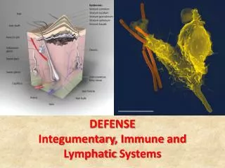
Integumentary Systems. Structure & Functions of Integumentary System Integumentary system consists of layers of the four types of body tissues: 1) epithelial tissue - outer layer of skin 2) connective tissue - tough & flexible protein fibers that act to hold body together

E N D
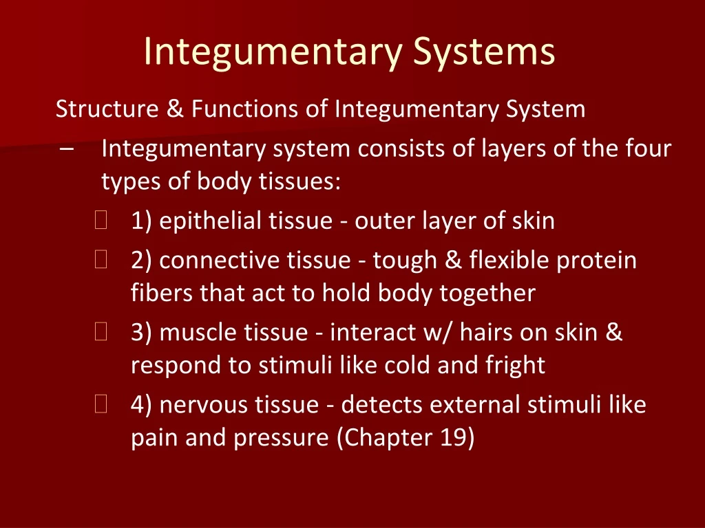
Integumentary Systems Structure & Functions of Integumentary System • Integumentary system consists of layers of the four types of body tissues: • 1) epithelial tissue - outer layer of skin • 2) connective tissue - tough & flexible protein fibers that act to hold body together • 3) muscle tissue - interact w/ hairs on skin & respond to stimuli like cold and fright • 4) nervous tissue - detects external stimuli like pain and pressure (Chapter 19)
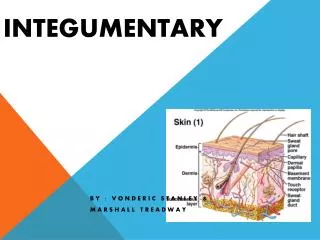
Skin: The Body’s Protection • Main organ in integumentary system is the skin, which makes it the largest organ in the body since it is 12-15% of body weight! • Made of two layers: • Epidermis: outer layer of skin • Dermis: inner layer of skin
Epidermis Outermost layer of skin made of 2 parts: exterior and interior portions • Exterior: 25-30 layers of dead, flattened cells that are continually being shed • Even though dead, are important since contain keratin which helps protect living cells underneath
Epidermis • Interior: living cells that continually divide to replace dead cells • Contain pigment melanin that colors skin and protects it from damage by solar radiation • Melanin is not sole protector for sun damage – can get skin cancer if are dark pigmented! • process of shedding takes 28 days (4 weeks)
Dermis • Inner, thicker portion of skin • Contains many structures: • Blood vessels (arteries & veins) • Nerves & nerve endings • Hair follicles • Sweat glands • Sebaceous (oil) glands • Muscles (to make hair stand up)
Subcutaneous Layer • Beneath dermis is subcutaneous layer • Made of fat and connective tissue • Help body absorb impacts, retain heat, store food
Functions of Skin • 1. Maintains homeostasis • Regulates internal body temperature • When temperature rises, small blood vessels in dermis dilate (increase in circumference), allowing blood flow to increase, so blood loses heat • When temperature lowers, blood vessels constrict (decrease in circumference), decreasing blood flow, so blood keeps in heat Feedback loop: Backward/forward
- + Feedback (Homeostasis) Loop Internal Body Temperature Changes Blood vessels dilate Blood vessels constrict Blood flow increases Blood flow decreases Blood loses heat Blood keeps in heat Internal Body Temperature Normalizes
2. sensory organ • Nerve cells get information from external environment about pain, pressure, and temperature and send message to brain • 3. produces Vitamin D • When exposed to UV light, skin makes Vitamin D, which is essential to help body absorb calcium • Most calcium supplements contain Vitamin D for that same reason • 4. protective layer • Shields underlying tissues from physical and chemical damage and from invading pathogens (viruses and bacteria)
Skin injury and Healing • Injuries to skin can occur due to scrapes, cuts, or burns, but how skin heals depends on severity • Mild scrape (no blood, epidermis only) • Deepest layer of affected epidermal cells start to divide to fill in gap left by abrasion • Cut (blood, epidermis and dermis) • Blood flows out of wound until clot forms • Scab develops, creating barrier between bacteria on skin and underlying tissues
Skin injury and Healing • Bacteria already present in wound gets killed by white blood cells that migrate to site • New skin cells begin repairing wound from beneath • Scab ‘falls’ off when new skin is formed • Large wound needs high amount of connective tissue which may form a scar
Healing of a Cut Before Cut in skin Blood pools, creating scab Skin cells regenerate from bottom up
Skin Burns • Burn (Sun, chemicals, hot objects) • First degree (mild sunburn) • Death of epidermal cells • Redness and mild pain • Heal in 1 week w/out scar • Second degree • Damage of both epidermal and dermal cells • Blistering and scaring may occur
Skin Burns • Burn (sun, chemicals, hot objects) • Third degree • Destruction of both epidermal and dermal cells • Skin function is lost, so skin grafts are required • Fourth degree • Destruction through skin and into muscles, tendons, ligaments, and bone
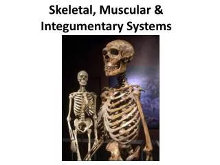
Bones: The Body’s Support Skeletal System Structure • Adult human skeleton contains 206 bones! Made of two main parts: • Axial skeleton: skull and bones that support it like vertebral column, ribs, sternum • Appendicular skeleton: bones of arms and legs (appendages), and all structures associated with them (shoulder, hips, wrists, ankles, fingers, toes)
Skeletal joints • Bones meet other bones at areas called joints • Joints facilitate movement of bones in relation to one another • Joints can be fixed (non-moveable) or non-fixed (moveable) • Fixed joints: skull
Skeletal joints • Non-fixed joints: knee, wrist, etc. - 4 types of moveable joints: * Ball-and-socket: hips, shoulders * Pivot: twisting arm at elbow * Hinge: elbows, knees, fingers, toes * Gliding: wrists, ankles
Ligaments • Joints are held together by ligaments • Ligament: tough band of connective tissue that attaches one bone to another • Joints with a large range of motion (knee) have many ligaments
Cartilage • Ends of bones are covered in cartilage • Allows for smooth movement between bone ends • Cushions joints
Bursae • Certain joints have fluid-filled sacs called bursae (bursa is singular) • Outside of joint between tendon and bone to reduce friction
Tendons • Muscles are attached to bones with tendons • Tendons are thick bands of connective tissue
BONE MUSCLE CARTILAGE JOINT LIGAMENT JOINTS TENDON
Types of Bone • Two types of bone tissue: • Compact bone and spongy bone • Compact bone: hardened bone that contains tubular structures called osteons (or Haversian systems) • Surrounds spongy bone to protect it • Spongy (cancellous) bone: less dense bone with many holes and spaces • Living bone cells are called osteocytes, which receive oxygen and nutrients from small blood vessels
Formation of Bone • Skeleton of human embryo is actually made of cartilage, not bone (same as what nose is made of) • Not until embryo is 9 weeks does cartilage get replaced by bone • When blood vessels penetrate cartilage membrane, stimulate it to become osteoblasts (precursors to osteocytes)
Human skeleton growth • Human skeleton is almost 100% bone, with cartilage found only in places where flexibility is needed – nose, ears, vertebral disks, and joint linings • Bone grows in length and diameter as result of sex hormones released during growth • Length: from cartilage plates at ends of bones • Diameter: from outer surface of bone • After growth stops, bone-forming cells are involved in repair and maintenance
Skeletal System Functions • Function of skeleton is five-fold: • 1. Provide framework for tissues of body • Allows muscles to attach to bones so they can provide movement to body • 2. Protects internal organs • 3. Produce blood cells • Red marrow: where red blood cells, white blood cells, blood clotting factors are produced • found in humerus, femur, sternum, ribs, vertebrae, pelvis
Skeletal System Functions • Function of skeleton is five-fold: • 4. Store fat • Yellow marrow: many other bones store fat in here • 5. Mineral storage • Body’s supply of calcium and phosphorous is stored in bone
Skeletal injury & disease • Skeleton is vulnerable to injury and disease • Broken bones • Too much force against bone can cause it to break or fracture • Physician must set bone back in place so new osteocytes may form in broken area and put two ends back together
Skeletal injury & disease • Skeleton is vulnerable to injury and disease • Osteoporosis • Loss of bone volume and mineral content which leads to bones becoming more porous and brittle and more susceptible for breakage • More common in older women since they produce lower amounts or estrogen which aids in bone formation
Osteoarthritis • Joints can become diseased • Arthritis: inflammation of the joints • Bone spurs are outgrowths of bone inside the joints so it limits mobility
Muscle Muscles • Nearly half of body mass is muscle! • Muscle: groups of fibers, or cells, bound together. Almost all muscle fibers have been present since birth • 3 main types of muscle: • Smooth muscle: walls of internal organs and blood vessels • Cardiac muscle: heart muscle • Skeletal muscle: muscles attached to bones
Smooth Muscle • Made up of sheets of cells that form a lining for organs • Most common function is to squeeze via contractions, exerting pressure on space inside tube or organ to move material inside it • Ex: food bolus gets squeezed through digestive system until it comes out; semen gets squeezed through vas deferens, then urethra
Contraction (AKA peristalsis) Item to be moved Movement of Smooth Muscle Smooth muscle of vessel or organ Contractions are involuntary (can’t be controlled by human) so smooth muscle is considered to be an involuntary muscle Direction of movement
Cardiac Muscle • Found in heart and is adapted to generate and conduct electrical impulses! • Considered an involuntary muscle
Skeletal Muscle • Muscle that is attached to and moves bones • Makes up majority of muscles in body which work in opposing pairs • Muscle X on one side of bone, Muscle Y on other side of bone • If Muscle X is contracted, Muscle Y is relaxed, and vice versa • Considered a voluntary muscle since contractions can be controlled • How do we contract our muscles?
Opposing Muscle Pairs Muscle Contracted Muscle Relaxed
Skeletal Muscle Contraction • All muscle tissue is made of muscle fibers, which are very long, fused muscle cells • Each fiber is made of smaller units called myofibrils • Myofibrils made of thick and thin filaments • Thick filaments: myosin • Thin filaments: actin • Myofibril can be divided into segments called sarcomeres
Muscle Contraction Relaxed Sarcomere • How do muscles contract? How do they know that you want to “make a muscle?” • Sliding Filament Theory Z Disc Actin Myosin