Download

1 / 11
130 likes | 203 Views
Explore the physical principles, hyperfine coupling, disorder analysis, rotational mobility, and site-directed spin labeling using EPR techniques for biophysical studies. Learn about detection methods and applications in determining structural information.

E N D
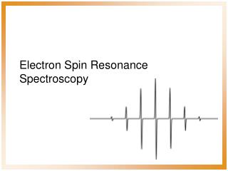
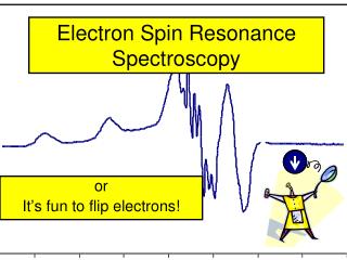
Electron Paramagnetic Resonance Biophysical Tools '02 - EPR
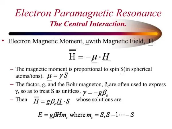
S I +1 0 -1 +½ hn hn E -½ -1 0 +1 Ho Physical principle Hyperfine coupling Biophysical Tools '02 - EPR
H C 3 O O O S N O S RLC cys154 N N ELC cys177 cysteine O O RLC cys125 M S L M T T S L cys707 Instrument amplifier klystron detector diode resonator magnet Biophysical Tools '02 - EPR
disorder • • average angle disorder Ÿ Ÿ z H x || o O N x y H y || o H z || o average angle Anisotropy and orientation width splitting Biophysical Tools '02 - EPR
Rotational mobility tr = 20 nsec tr = 200 msec tr = 10 nsec tr = 23 msec tr = 4 msec tr = 0.001 nsec 4 Biophysical Tools '02 - EPR tr = 0.001 nsec tr = 0.001 nsec tr = 0.001 nsec tr = 0.001 nsec
force / movement Head Orientation and Mobility in ATPase cycle • sequential transitions from disordered-mobile myosin heads to ordered-immobile heads drives the generation of force Biophysical Tools '02 - EPR
Site Directed Spin Labeling EPR Hubbell, 1989 “cysteine scanning” from 130-146
4 P1/2(O2)/ P1/2(CROX) P1/2(O2)/ P1/2(CROX) P1/2= 60 mW amplitude 2 P1/2= 20 mW 0 0 5 10 15 power ½(mW) ½ Secondary structure determination Biophysical Tools '02 - EPR
Rabenstein & Shin, PNAS, 92 (1995) Range: 8-20 Å Dipolar EPR: distances Non-interacting spins Double labeled
Spin trapping Biophysical Tools '02 - EPR
Transition metals Biophysical Tools '02 - EPR