Respiratory system-part 1
200 likes | 411 Views
Respiratory system-part 1 . Marilyn Rose RT, RDMS. Outline Part 1. Physiology of Respiratory System Internal devices Congenital Hereditary Inflammatory Diffuse Lung disease. Physiology. Role= oxygenation of blood and removal of carbon dioxide

Respiratory system-part 1
E N D
Presentation Transcript
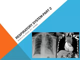
Respiratory system-part 1 Marilyn Rose RT, RDMS
Outline Part 1 • Physiology of Respiratory System • Internal devices • Congenital Hereditary • Inflammatory • Diffuse Lung disease
Physiology • Role= oxygenation of blood and removal of carbon dioxide • Upper- outside thorax- nasopharynx, oropharynx, larynx- passage of air into lower system • Lower- within thoracic cavity- trachea, bronchi and bronchioles • Trachea branches into two bronchi at the carina • Tracheobronchial tree- lined with mucous membrane- cilia • Gas exchange • external respiration- at alveoli- (cluster- acinus) parenchyma of lung • internal respiration- O2 from alveoli- blood capillaries attach to Hb and circulates to the body tissues.
Respiration • CO2 – waste product of cell metabolism- opposite direction- blood capillaries, alveoli and exit body at expiration. • - CO2 regulate the respiratory center (medulla) to stimulate diaphragm/ intercostals to contract- decreased lung pressure forces air out. • Lung has two blood supplies- • pulmonary circulation-low pressure/ low resistance- O2 enter CO2 exit • bronchial circulation- high- O2 blood to lung • Double walled membrane • 2 layers of pleura- • visceral-inner- adhere to lung • parietal- inner chest wall • Potential space between pleura – surfactant • fluid can collect from inflammatory/neoplastic process
Internal device • Endotracheal tube • crx always taken post placement- ensure proper placement 5-7 cm above carina (flexion/ extension will move 2 cm cranial/ caudal) • too low? Extend in Rt bronchus- cause lt lung atelectasis • too high? Gastric dilation- regurgitation/ aspiration pneumonia • Central venous catheters • inserted into subclavian VEIN • optimal location of tip is where brachiocephalic veins join- SVC • xray- medial to anterior border of first rib • measure central venous pressure • conduit for rapid transfusion • Swan-Ganz– measures cardiac output- placed in the pulmonary artery- radiopaque strip • Transvenous Cardiac Pacemakers • maintain cardiac rhythm with heart block or bradyarrhythmias positioned at the apex of Rt ventricle
Congenital/ hereditary diseases • Cystic fibrosis • excessive viscous mucus by exocrine glands • Defective gene on chromosome 7 • Most common among white children • 90% morbidity rate from respiratory • CF affects pancreas/ GI/ and Liver • Hyaline Membrane Disease • Idiopathic respiratory distress syndrome • most common cause of respiratory distress in newborn (premature) • Underaeration of lungs due to lack of surfacant • Radiographic- • Ground glass appearance of parenchyma • air bronchogram • Treatment- artificial surfactant and + pressure ventilator
Cystic fibrosis • Radiographic- • Thick hilar markings- serial CT • Treatment • Prophylactic antibiotics • Chest physiotherapy • bronchodilators • Lungs • Mucous plugs lead to focal area of lung collapse • Common recurrent pulmonary infections • abdomen • Digestion of fat impaired by blockage of ducts at pancreas • prevent digestive enzymes from entering duodenum • skin • Sweat glands- excessive perspiration= “sweat test”
Inflammatory disorders- upper • Croup • Viral infection of young children • Inflammatory obstructive swelling at subglottic trachea • Causes- stridor or barking cough • Radiographic- soft tissue neck x-ray= hourglass shape from edema • Treatment- cool mist/ steam from a hot shower • Epiglottitis • Acute infection caused by haemophilus influenzae • Cause thick epiglottic tissue • Decreased from the vaccine for type B (HiB) routinely used • Radiographic- epiglottis looks like an adult thumb • The condition can result in complete airway obstruction
Inflammatory disorders- lower • Pneumonia • Anthrax • Lung abscess • TB • Pulmonary Mycosis • RSV • SARS bacterial viral TB
pneumonia • inflammation of lung by bacteria or viruses • Alveolar- • inflam exudate replaces air- affected lung solid • consolidation of parenchyma- air bronchogram sign • no airway obstruction • Bronchopneumonia- • small patches of consolidation- • airway obstruction- atelectasis- no air bronchogram • scattered opacities • Interstitial • Viral- affects walls and lining of alveoli • linear or honeycomb lung • Aspiration • Aspirate esophageal or gastric contents- • general anesthesia risk- • multiple alveolar densities- posterior segments of up/ lower lobes are most affected alveolar interstitial aspiration
anthrax • Caused by a sporelike microbe • biologic terrorist attacks in 2001 caused its return • Cutaneous, inhalation and gastrointestinal routes of entry • Cutaneous- most common- working with animal hides • GI route causes mesenteric adenopathy • Inhaled anthracis is most lethal – • germinating in lung and lymph nodes • Mediastinal widening • Pleural effussions (w/o infiltrates)
Lung abscess • Necrotic area of pulmonary parenchyma containing purulent material • Can be complicated by pneumonia, bronchobst, aspiration, foreign body or septic embolism • Clinically- fever and cough - copious sputum • Complication is a brain abscess • On xray a spherical density with a hazy border- can develop into an air/ fluid level
TB • Mycobacterium tuberculosis- • bacteria- wax coat- live outside the body for long time • Spreads by droplets in the air- sometimes by dried sputum inhalation • Primarily disease of the lungs but can involve the GI and skeletal system • Initial infection- • inflam cells collect around bacteria form a tubercle- • depending on number and resistance for the outcome • Good resistance • fibrous tissue surrounds “tumor” prevents spread- scar in the apical segment / calcium deposits • The core becomes necrotic- then a liquefied cavity (air/fluid level) • low resistnce • diffuse distruction throughout the lung- huge cavities and fatal
Types of TB • Primary TB- • lobar, air-space consolidation, dense, well defined, hilar nodes, pleural effussion- apical lordotic position is used • Miliary TB- • dissemination of the disease by the bloodstream- nodules distributed to both lungs • Secondary TB- • reactivation from dormant tubercles- decrease in immune system- hazy radiating form the hilum of lung • Tuberculoma- • sharp circumscribed parenchymal nodule that can develop primary or secondary- break down anytime and disseminate the disease
Pulmonary mycosis • Fungal infection of the lung • Histoplasmosis (Mississippi River Valley/ Ohio River) • Radiographic appearance = similar to TB- but benign • Fibrosis in mediastinum • Can cause obstruction- • svc, pulm art/ veins, narrow esophc • Ca++ of liver, spleen, and lymph nodes- diagnostic!!! • Coccidioidomycosis( Desert of SW United States) • Spread by fungal spore in air • Symptoms similar to influenzae • Chronic dx= lung abscess • Mult areas of pulm infiltration- lower lung- hilar lymph node • Lymph node Ca++ = popcorn • 3cm low lobe with ca++ w/in mass
RSV and SARS • Respiratory syncytial virus • Affect almost all children by age 2 • Fewer than 2% need to be hospitalized • Lower respiratory infection • necrosis of epithelium- bronchiolitis • Obstruction, interstitial pneumonia • Radiographic- hyperinflation, >interstitial markings • SARS= severe acute respiratory syndrome • Global 2003- 1st in China • Droplet- upper and lower infection • Cough, hypoxemia, fatal 3% • Radiograph- lungs appear normal • progress to patchy infiltrates- consolidation
Diffuse lung disease • COPD • Chronic obstruction of airway- two disease processes coexist • Chronic bronchitis- excessive mucus- leads to obstruction of airway • 90% cigarette smoke, sever coughing and sputum production- • narrow airways and over inflation of lungs • Radiographic- • dirty chest- increased bronchovascular markings • Emphysema- distention of air spaces- destroy alveolar walls- asthma • Result is bronchiectasis- chronic dilation of bronchi • Destructive changes in small airways lead to increased volume of air in lungs • Radiographic- • flattening of domes of diaphragm, > retrosternal air, barrel chest, >vascular markings • Bullae- air containing cystic spaces • Cause- cigarette smoke, air pollution, occupational exposure
Asthma and Bronchiectasis • Asthma • Widespread narrowing of the airways due to > responsiveness to allergens • Extrinsic- dust, pollen, animal dander, foods • Intrinsic- exercise, heat/ cold, emotional upset • Radiographic- • acute= no abnormalities • Chronic= dirty chest with normal vascular markings • Bronchiectasis • Permanent abnormal dilation of one or more large bronchi • Destroy elastic walls • Recurrent episodes of pneumonia/ hemopysis • Radiographic- circular cystic spaces or honeycomb
Sarcoidosis • Multisystem granulomatous disease • Young adults • Multiple epithelioid granulomas • Radiographic- usually found on screening CXR • Symmetric hilar lymph node enlargement, diffuse interstitial pattern • Skeletal lesions in small bones of hands and feet • Some patients have elevated serum calcium= lead to nephroca++
Pneumoconiosis • Prolonged occupational exposure to irritating particulates • These inhaled foreign substances • malignant neoplasm • Severity depends on size, length of exposure, concentration in atmosphere • Silicosis- most common • inhalation of silicon dioxide- mining, foundry and sandblasting or quartz dust • Rad- eggshell at periphery • Asbestosis- • Improperly protected workers engaged in handling building materials, insulation composed of asbestos • Major complication= mesothelioma- malignant pleural tumor • Rad-ca++ of linear pleural plaques • Anthracosis • Coal miner- working with anthracite- hard coal • Black lung • Rad- hyperinflated and homogeneous mass at hilar • region