Download
1 / 27
2.83k likes | 9.64k Views
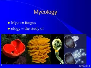
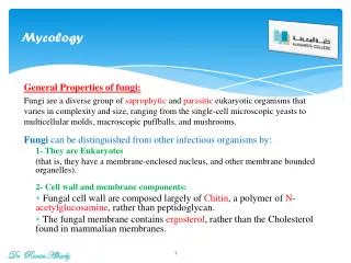
INTRODUCTION TO MYCOLOGY. By. Dr. Emad AbdElhameed Morad. Lecturer of Medical Microbiology and Immunology. Fungi are eukaryotic organisms. Their cell wall consists of chitin . Their cell membrane contains ergosterol . Classification. Morphological. Clinical. Systematic.

E N D
INTRODUCTION TO MYCOLOGY By Dr. Emad AbdElhameed Morad Lecturer of Medical Microbiology and Immunology
Fungi are eukaryotic organisms. • Their cell wall consists of chitin. • Their cell membrane contains ergosterol. Dr. Emad AbdElhameed Morad
Classification Dr. Emad AbdElhameed Morad
Morphological Clinical Systematic Dr. Emad AbdElhameed Morad
Fungal morphology Yeast Mold Dimorphic Dr. Emad AbdElhameed Morad
Yeasts • Oval or round cells that reproduce by budding to form blastospores. • May form pseudohyphae(if blastospores remain attached to each other). • Examples: Candida, Cryptococcus. Dr. Emad AbdElhameed Morad
Budding yeast cells Pseudohyphae Dr. Emad AbdElhameed Morad
Molds • Also called filamentous fungi or mycelial fungi. • Formed of filaments called hyphae. • Hyphae interlace to form mycelium. • Hyphae may be septate or aseptate. • Reproduce by formation of conidia. • Conidia may be unicellular (microconidia) or multicellular (macroconidia). • Examples are: dermatophytes & aspergillus. Dr. Emad AbdElhameed Morad
Hyphae Mycelium Microconidia Macroconidia Dr. Emad AbdElhameed Morad
Dimorphic fungi • These fungi occur in two forms: • At the room temperature (22 degree), it appears as mold. • In the body (37 degree), it appears as yeast cells. • Examples: Histoplasma & Blastomyces. At 22 degree At 37 degree Dr. Emad AbdElhameed Morad
Clinical classification Superficial mycoses Cutaneous mycoses Subcutaneous mycoses Systemic mycoses Opportunistic mycoses Allergy & mycetismus & mycotoxicosis Dr. Emad AbdElhameed Morad
Superficial mycoses • Fungal infections confined to the stratum corneum without tissue invasion. • Example: Tinea versicolor caused by Malassezia furfur. Dr. Emad AbdElhameed Morad
Cutaneous mycoses • Fungal infections that involve keratinized tissues as skin, hair, nail. • Example: Tinea caused by dermatophytes. Dr. Emad AbdElhameed Morad
Subcutaneous mycoses • Fungal infections that are confined to subcutaneous tissues without dissemination to distant sites. • Example: mycetoma (madura foot). Dr. Emad AbdElhameed Morad
Systemic mycoses • Also called endemic mycoses. • Begin as primary pulmonary lesions that may disseminate to any organ. • Caused by dimorphic fungi. Dr. Emad AbdElhameed Morad
Opportunistic mycoses • Affect immunocompromised individuals • Examples are: • Candidiasis caused by Candida albicans. • Cryptococcosis caused by Cryptococcus neoformans. • Aspergillosis caused by aspergillus fungus. • Pneumocystis pneumonia caused by pneumocystis jiroveci in AIDS patients. Dr. Emad AbdElhameed Morad
Allergy • Allergy occurs to fungal spores particularly those of aspergillus fungus. • Example: bronchial asthma. • The fungal flesh itself is toxic. • Example: Amanita mushroom poisoning. Mycetismus Dr. Emad AbdElhameed Morad
Mycotoxicosis • Example: Aflatoxins produced by Aspergillus flavus which infects grains and peanuts. This toxin is hepatotoxic and cause tumors in animals and suspected of causing hepatic carcinoma in humans. Dr. Emad AbdElhameed Morad
Systematic classification • It is based on the type of fungal spores: • Sexual spores • Asexual spores Dr. Emad AbdElhameed Morad
Sexual spores • Zygospores: • Fungi forming zygospores are called zygomycetes. • Ascospores: • Ascospores are carried in ascus. • Fungi forming ascospores are called ascomycetes. • Basidiospores: • Basidiospores are carried on basidium. • Fungi forming basidiospores are called basidiomycetes. Deuteromycetes are fungi whose sexual spores are unknown. But, they produce asexual spores. Dr. Emad AbdElhameed Morad
Zygospores Ascospores Basidiospores Dr. Emad AbdElhameed Morad
Asexual spores • Blastospores: • Produced by budding of the yeast cells. • Conidia: • Produced by molds. • May be microconidia or macroconidia. • Arthrospores: • Produced by fragmentation of hyphae. • Chlamydospores: • Rounded thick walled spores produced by candida fungus. Dr. Emad AbdElhameed Morad
Blastospores Microconidia Macroconidia Arthrospores Chlamydospores Dr. Emad AbdElhameed Morad
Antifungal drugs • Selective toxicity is very limited in antifungal drugs because fungi like human cells are eukaryotic. • They are: • Amphotericin B: • Binds to the ergosterol in the fungal cell membrane. • Used in severe fungal infections. • Nephrotoxic. • Flucytosine: • Inhibit fungal DNA synthesis. Dr. Emad AbdElhameed Morad
Azoles: • Like ketoconazle, fluconazole, itraconazole. • Inhibit ergosterol synthesis. • Terbinafine: • Inhibit ergosterol synthesis. • Used in treatment of dermatophytes. • Griseofulvin: • Used in treatment of dermatophytes because it is concentrated in the keratinized tissues. • Topical antifungal agents: • Nystatin. • Clotrimazole and miconazole. Dr. Emad AbdElhameed Morad
Caspofungin: • New antifungal drug • Inhibit fungal cell wall chitin synthesis • Used in invasive fungal infections
GOOD LUCK Dr. Emad AbdElhameed Morad