Download
1 / 69
690 likes | 793 Views
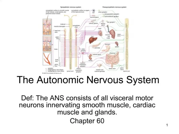
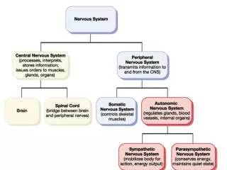
The Autonomic Nervous System. Autonomic Nervous System (ANS). The ANS consists of motor neurons that: Innervate smooth and cardiac muscle and glands Make adjustments to ensure optimal support for body activities Operate via subconscious control Have viscera as most of their effectors.

E N D
Autonomic Nervous System (ANS) • The ANS consists of motor neurons that: • Innervate smooth and cardiac muscle and glands • Make adjustments to ensure optimal support for body activities • Operate via subconscious control • Have viscera as most of their effectors
Divisions of the ANS • Sympathetic division (thoracolumbar, “fight or flight”) • Thoracic and lumbar segments • Parasympathetic division (craniosacral, “rest and repose”) • Preganglionic fibers leaving the brain and sacral segments • Enteric nervous system (ENS) • May work independently
Sympathetic and Parasympathetic • Often they have opposing effects • May work independently • May work together each one controlling one stage of the process
ANS Versus Somatic Nervous System (SNS) • The ANS differs from the SNS in the following three areas • Effectors • Efferent pathways • Target organ responses
Effectors • The effectors of the SNS are skeletal muscles • The effectors of the ANS are cardiac muscle, smooth muscle, and glands
Efferent Pathways • Heavily myelinated axons of the somatic motor neurons extend from the CNS to the effector • Axons of the ANS are a two-neuron chain • The preganglionic (first) neuron has a lightly myelinated axon • The ganglionic (second) neuron extends to an effector organ
Neurotransmitter Effects • All somatic motor neurons release Acetylcholine (ACh), which has an excitatory effect • In the ANS: • Preganglionic fibers release ACh • Postganglionic fibers release norepinephrine or ACh and the effect is either stimulatory or inhibitory • ANS effect on the target organ is dependent upon the neurotransmitter released and the receptor type of the effector
Sympathetic division anatomy • Preganglionic neurons between segments T1 and L2 – lateral gray horn of spinal cord • Preganglionic fibers • Short • Travel in the ventral root and spinal nerve • Ganglionic neurons in ganglia near vertebral column • Specialized neurons in adrenal glands • Postganglionic fibers • Long fibers
Sympathetic ganglia • Sympathetic chain ganglia (paravertebral ganglia) • Collateral ganglia (prevertebral ganglia) • Adrenal medulla
Organization and anatomy of the sympathetic division • Segments T1-L2, ventral roots give rise to myelinated white ramus • Leads to sympathetic chain ganglia
Postganglionic fibers of the sympathetic ganglia • Some fibers will return to the spinal nerve through a gray ramus and will innervate skin, blood vessels, sweat glands, adipose tissue, arrector pili muscle (body wall structures) • Postganglionic fibers coming from chain ganglia will form sympathetic nerves that will innervate thoracic organs
Collateral ganglia • Preganglionic fibers will pass through the sympathetic chain without synapsing • Preganglionic fibers will synapse within collateral ganglia • Preganglionic fibers synapsing within collateral ganglia will from Splanchnic nerves
Collateral ganglia • Celiac ganglion • Postganglionic fibers innervates stomach, liver, gall bladder, pancreas, spleen • Superior mesenteric ganglion • Postganglionic fibers innervates small intestine
Collateral ganglia • Inferior mesenteric ganglion • Postganglionic fibers innervate the large intestine • Inferior hypogastric • Postganglionic fibers innervates urinary bladder , sex organs
Adrenal medulla • Preganglionic fibers will pass through sympathetic chain ganglia and collateral ganglia without synapsing • Preganglionic fibers will then synapse on adrenal medulla • Adrenal medulla will secrete • Epinephrine • Norepinephrine
Adrenal medulla • Neurotransmitter will go into general circulation • Their effects last longer than those produced by direct sympathetic innervation
Role of the Sympathetic Division • The sympathetic division is the “fight-or-flight” system • Involves E activities – exercise, emergency • Promotes adjustments during exercise • Blood flow to organs is reduced, flow to muscles is increased
Role of the Sympathetic Division • Its activity is illustrated by a person who is threatened • Heart rate increases, and breathing is rapid and deep • The skin is cold and sweaty, and the pupils dilate
Parasympathetic division (craniosacral division) • Preganglionic neurons in the brainstem(nuclei of cranial nerves III, VII, IX, X) and sacral segments of spinal cord (S2-S4) • Ganglionic neurons in peripheral ganglia located within or near target organs • Terminal ganglion • Intramural ganglion
Organization and anatomy of the parasympathetic division • Preganglionic fibers leave the brain as cranial nerves III, VII, IX, X • Cranial nerve X provides 75% of the parasympathetic outflow • Sacral neurons form the pelvic nerves
Parasympathetic activation • Effects produced by the parasympathetic division • relaxation • food processing • energy absorption • Pupil constriction • Constriction of respiratory passageway • Decrease heart rate and blood pressure • Stimulates defecation and urination
Summary: The Anatomical Differences between the Sympathetic and Parasympathetic Divisions
Sensory Visceral Neurons • Are found in: • Sensory ganglia of cranial nerves • Dorsal root ganglia • Sympathetic ganglia • Afferent visceral fibers are found in: • Cranial nerves VII, IX, X • Autonomic nerves • Spinal nerves
Visceral Reflexes • Visceral reflexes have the same elements as somatic reflexes • They are always polysynaptic pathways
Referred Pain • Pain stimuli arising from the viscera are perceived as somatic in origin • This may be due to the fact that visceral pain afferents travel along the same pathways as somatic pain fibers
Neurotransmitters and Receptors • Acetylcholine (ACh) and norepinephrine (NE) are the two major neurotransmitters of the ANS • ACh is released by all preganglionic axons and all parasympathetic postganglionic axons • Cholinergic fibers – ACh-releasing fibers
Neurotransmitters and Receptors • Adrenergic fibers – sympathetic postganglionic axons that release NE • Neurotransmitter effects can be excitatory or inhibitory depending upon the receptor type
Neurotransmitters and parasympathetic functions • All parasympathetic fibers release ACh • Short-lived response as ACh is broken down by AChE and tissue cholinesterase • Postsynaptic membranes have two kinds of receptors: muscarinic and nicotinic
Neurotransmitters and parasympathetic functions • Muscarinic • Parasympathetic target organs • Postganglionic cholinergic fibers • Cardiac muscle • Smooth muscle • Excitatory or inhibitory effects • Depends on the receptor type of the target organ
Nicotinic Receptors • Nicotinic receptors are found on: • Surface of skeletal muscles • All ganglionic neurons of both sympathetic and parasympathetic divisions • Ganglionic neurons of the adrenal medulla • The effect of ACh binding to nicotinic receptors is always stimulatory by opening Na channels
Adrenergic Receptors • The two types of adrenergic receptors are alpha and beta • Each type has two or three subclasses (1, 2, 1, 2 , 3)
Adrenergic Receptors • Alpha 1 • Constrict blood vessels of: skin, mucosa, abdominal viscera, kidney, salivary glands, etc. • Dilates pupil • Constrict involuntary sphincters • Excitatory