Download
1 / 52
600 likes | 1.34k Views
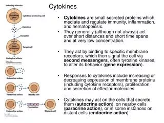
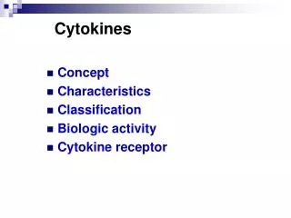
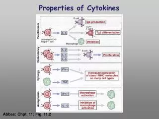
Properties of Cytokines. Abbas: Chpt. 11; Fig. 11.2. Cytokines, chemokines and growth factors can be placed into several structurally & functionally related families. Growth Factors ( direct hematopoiesis and endothelial cell growth/activity ) IL-1 Family (e.g., IL-1 & “Toll-like”)

E N D
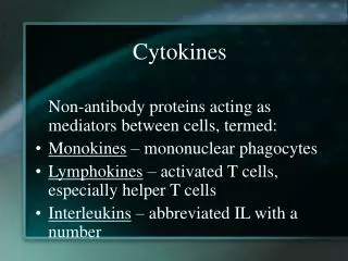
Properties of Cytokines Abbas: Chpt. 11; Fig. 11.2
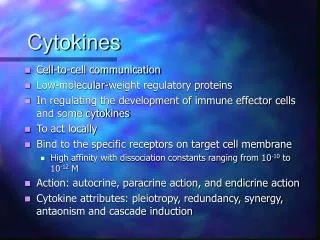
Cytokines, chemokines and growth factors can be placed into several structurally & functionally related families • Growth Factors (direct hematopoiesis and endothelial cell growth/activity) • IL-1 Family (e.g., IL-1 & “Toll-like”) • TNF Family (e.g., TNF-a, CD40L, FasL) • TGF-b Family (e.g., TGF-b ) • Chemokines (e.g., CC and CXC families) • Hematopoietins / a.k.a. Four Helix Bundle (e.g., IL-2, IL-4, IL-6, IL-10, IL-12, IFN-g, IFN-a/b)
Let’s digress to review TCR signaling for an important clinical pearl!
TCR-mediated Signal Transduction: A Tyrosine Kinase Cascade Abbas & Lichtman, Fig. 8-7, p. 175
NF-AT and TCR-mediated Signal Transduction Cyclosporin A (CyA) & Tacrolimus (FK506) are two important drugs that block calcineurin activation NF-AT activation IL-2 production! They are therefore potent immunosuppressive drugs. Abbas & Lichtman, Fig. 8-12, p. 183 (see Fig. 14.5 in Janeway, p618)
IL-4, IL-5 and IL-6 are Th2 cytokines and promote humoral immunity
Pathophysiology of the balance between Th1 and Th2 - Defense against virus & intra- cellular pathogens - Anti-tumor immunity DTH - Defense against parasites - Ab production & class switch Th1 Th2 Rheumatoid arthritis Type I Diabetes mellitus Multiple sclerosis Allergy Graft-vs-host disease
Functions of Complement A. Host Defense B. Disposal of Waste C. Regulation of the Immune Response
Functions of Complement Disposal of Waste Immune Complex Removal Apoptotic Cell Debris Removal
Phagocytosis: An Evolutionarily Conserved Mechanism to Remove Apoptotic Bodies and Microbial Pathogens
From: Lekstrom-Himes and Gallin, N Engl J Med, 343:1703, 2000
Clearance of pathogens Death of pathogenic microbe Persistence of pathogenic microbe Resolution of infection Failure of resolution of infection Clearance of apoptotic corpses Suppression of inflammation Inappropriate inflammation Tolerance Break in tolerance Immunological Consequences of Phagocytosis
Activating FcgR Inhibitory FcgR g g Syk SHIP ITAM ITIM + - Phagocytosis
Requirement of Activating FcgRs in Immune Complex-mediated Glomerulonephritis Strain: C57Bl/6 NZB/NZW NZB/NZW g chain: -/- -/- +/- Glomerulonephritis is blocked in g chain-deficient NZB/NZW (lupus-prone) mice. Pathological features include mesangial thickening and hypercellularity evolving into end-stage sclerotic and crescentic changes. From: Clynes et al., Science 279:1052, 1998.
Summary Phagocytosis is a component of innate and aquired immunity. It is the principal means of destroying pathogenic bacteria and fungi. Phagocytosis initiates the process of antigen presentation. Many phagocytic receptors recognize a diverse array of microbial pathogens. Some pathogens (e.g., S. pneumoniae) require opsonization for their clearance. Bugs fight back. Phagocytosis is an essential component of development and tissue remodeling. Ingestion of apoptotic bodies is immunologically “silent” and is normally accompanied by a suppression of inflammation. Failure of this mechanism may result in autoimmunity. Fc receptors come in two basic types: activating (ITAM-associated) and inhibitory (ITIM-associated). The relative expression of activating and inhibitory Fc receptors determines the outcome of a given engagement of Fc receptors. Fc receptor-driven pathology includes formation and deposition of immune complexes, which play a major role in autoimmunity.
Receptors Important in The Systemic Response to Infection
TLR Signaling Components Vertebrates Drosophila TLR-4 Toll CD14 Receptor Complex MD2 extracellular space cytosol TIR domain MyD88 Tube Adaptor proteins Kinases DD DD IRAK Pelle ECSIT dECSIT TRAF6 dTRAF TAK MEKK1 Ird6 IKK-g IKK complex IKK-a IKK-b TIR = Toll/IL-1 receptor DD = Death domain IKK = I-kB kinase Cactus I-kB NF-kB p50 p65 Dif/Relish
The (Primary) Acquired Immune Response is Initiated by Innate Immune Recognition
Chemokines Direct Trafficking of Immune Cells From: Luster, Curr. Opin. Immunol. 14:129, 2002
T APC Autoimmune diseases: classification according to the class of the susceptibility MHC allotype and lineage of autoantigen specific T-cells mediating injury Class II Class I CD8 CD4 HLA-A,B, or C HLA-DR, DQ, or DP Psoriasis Psoriatic arthritis Reiter’s syndrome Ankylosing spondylitis Multiple sclerosis Pemphigus vulgaris Rheumatoid arthritis Lupus erythematosus
Stages to progression of autoimmune disease Genetic Predisposition MHC allele (+ other genes?) Initiation of Immune Recognition Event (+ other genes?) T cell clonal Expansion, Spreading B cell help Effector mechanisms Bind self peptides Select latently autoreactive TCR repertoire Autoimmune Disease Autoimmunity (Years) Inciting event /failure of tolerizing mechanism
Stages to progression of autoimmune disease Genetic Predisposition MHC allele (+ other genes?) Initiation of Immune Recognition Event (+ other genes?) T cell clonal Expansion, Spreading B cell help Effector mechanisms Bind self peptides Select latently autoreactive TCR repertoire Autoimmune Disease Autoimmunity (Years) Inciting event /failure of tolerizing mechanism
Hyperacute Minutes to hours Preexisting antibodies (IgG) Intravascular thrombosis Hx of blood transfusion, transplantation or multiple pregnancies Acute Rejection Few days to weeks CD4 + CD8 T-Cells Humoral antibody response Parenchymal damage & Inflammation Chronic Rejection Chronic fibrosis Accelerated arteriosclerosis 6 months to yrs CD4, CD8, (Th2) Macrophages Not Applicable Primary Graft Failure 10 – 30 Days Host NK Cells Lysis of donor stem cells Secondary Graft Failure 30 days – 6 months Autologous T-Cells CD4 + CD8 - Lysis of donor stem cells Pathological Mechanism of Rejection Solid Organ Bone Marrow/PBSC
ORAL TOLERANCE • ORAL ADMINISTRATION OF A PROTEIN ANTIGEN MAY LEAD TO SUPPRESSION OF SYSTEMIC HUMORAL AND CELL-MEDIATED IMMUNE RESPONSES TO IMMUNIZATION WITH THE SAME ANTIGEN. • POSSIBLE MECHANISMS: • INDUCTION OF ANERGY OF ANTIGEN-SPECIFIC T CELLS • CLONAL DELETION OF ANTIGEN-SPECIFIC T CELLS • SELECTIVE EXPANSION OF CELLS PRODUCING IMMUNOSUPPRESSIVE CYTOKINES (IL-4, IL-10, TGF-b)
REGULATORY T CELLS(CD4+) • TH3 CELLS: A POPULATION OF CD4+T CELLS THAT PRODUCE TGF-b. ISOLATED FROM MICE FED LOW DOSE OF ANTIGEN FOR TOLERANCE INDUCTION • TR1 CELLS: A POPULATION OF CD4+T CELLS THAT PRODUCE IL-10. CAN PRODUCE SUPPRESSION OF EXPERIMENTAL COLITIS IN MICE • CD4+CD25+ REGULATORY T CELLS: A POPULATION OF CD4+T CELLS THAT CAN PREVENT AUTOREACTIVITY IN VIVO.
INDUCTIVE LYMPHOEPITHELIAL TISSUES:PEYER’S PATCHES M CELLS B B APC T B ACTIVATED LYMPHOID FOLLICLE B T B T B T PERIPHERAL BLOOD MESENTERIC LYMPH NODES THORACIC DUCT
EFFECTOR SITES: LAMINA PROPRIA AND INTRAEPITHELIUM DISTANT GUT MUCOSA T8 T8 T4 APC T4 SC SIgA IgA-J B PERIPHERAL BLOOD OTHER EXOCRINE TISSUES
Inflammatory Bowel Disease: Immunological Features • HUMORAL IMMUNITY: MASSIVE INCREASE IN THE NUMBER OF PLASMA CELLS AND IN IgG PRODUCTION (IgG2 IN CD AND IgG1 IN UC) • IMBALANCE OF PRO-INFLAMMATORY (TNF-a, IL-1,IL-8, IL-12) AND ANTI-INFLAMMATORY CYTOKINES (IL-10, IL-4, IL-13)
Immune response to HIV-1 and effects of HIV infection CD4 T cells #/ml Flu-like Illness Asymptomatic phase Symptomatic phase CLINICAL AIDS Chronic lymphadenopathy Mucous membrane infections
R5 is almost always thesexually transmissible form of the virus Primary isolates from newly infected individuals are usually R5 R5 strainsmainly replicate in monocytes. Activated and memory T cells are infected, but at lower efficiency (old term = MT-tropic or monocytotropic) Therefore much of the viral load in earlier phase of HIV infection is in the monocytes and macrophages and the numbers of CD4 T cells remains stable, but decreased HIV strain early in infection
CD8 T-cell Response to HIV-1 Establishes asymptomatic phase of infection The CD8 T-cell responds to HIV-peptides by activation, clonal expansion, and differentiation to effector status Specific lysisof HIV- infectedtarget cells (macrophages and CD4 T cells) via perforin pathway and/ or apoptosis via upregulation of fas ligand Stronginhibition of viral infectivity by release of chemokines (MIP-1/, RANTES) that bind to CCR5 and block coreceptor dependent entry of R5 HIV-1 Release of IFN- and secondarily TNF-, decrease LTR-driven transcription
Thwarted immunosurveillance (2) Dendritic cells used as a “Trojan Horse” • Immature DCs, typically located in the submucosa express a C-type lectin DC-SIGN • HIV-1 envelope binds to DC-SIGN with high affinity • The virions are internalized and remain in acidic endosomal compartments while the DC matures • Intact infectious virions are reexpressed on the surface when the DC enters the lymph node
Viral Response near end of asymptomatic period Rate of viral infection and potential mutations increases. Definitive viral escape occurs when virus is no longer presented by MHC to available CD8 T cell clones Continual generation of env mutations Selection against R5 variants by CD8 T-cell CCR5 chemokines that blocks infectionis finally bypassed Change in cellular tropism by env mutations leads to X4 phenotype (CXCR4, T-tropic) Enhanced T-tropism of X4 leads to more significant impairment of CD4 T-cell compartment Loss of the “epitope war”
Another reason for CD4 T cell loss CD4 T cell activation initiates HIV replication HIV replication initiates CD4 T cell activation T cell activation causes, among other effects, a marked increase in cyclin T1, NFAT and NFkB This links viral expression to T cell activation
FcR EBV CR2 SmIg gp350/220 MHC II B Cell EBV Latency, Immortalization and the Role of T Cells FcR Endocytosis of the EBV-CR2 complex CR2 EBV genomes exist in latent form intracellularly as circular plasmids; also EBV genome can integrate into cellular genome Latent infection FcR CR2 MA Depressed T cell function Immortalization (Burkitt's Lymphoma) EBV genome EBNA
IgE-mediated InflammationEarly Phase Time course: Minutes after antigen challenge Example: Acute asthma Cause: Mediators released by cells attracted to area of inflammation Cells involved: Mast cells, basophils
IgE-mediated InflammationLate Phase Time course: Hours after antigen challenge Example: Chronic asthma Cause: Mediators released by cells attracted to area of inflammation during and after the early phase Cells involved: Eosinophils, Basophils Neutrophils, Lymphocytes
Control of IgE Production(Candidate Genes) I. Localization to specific chromosomes a. Chromosome 5q - Promoter variants for IL-4 (IL-3, -5, -9, -13 and GM-CSF) b. Chromosome 11q Subunit of FcRI (High affinity IgE receptor) c. Others II. HLA linkage to specific antigen responses
“Hygiene Hypothesis” • Observation (one of a number of examples) – Children raised in rural areas close to animals and exposed to endotoxin in dust have a lower incidence of atopic disease • Theory – Endotoxin acting on Toll-like receptors influences the cytokines that APC’s secrete as they present antigen so as to favor a Th1 instead of a Th2 response
Inflammatory Mediators Mast Cells and Basophils Histamine Leukotrienes C4, D4, E4 Platelet Activating Factor (PAF) TNF-a,IL-4, IL-13 Mast Cells Only PGD2 Tryptase (Used to detect anaphylaxis) IL-5, -6