Download

1 / 52

E N D
Cytokines in diseases M.Prasad Naidu MSc Medical Biochemistry, Ph.D,.
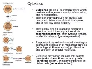
Introduction • Cytokines are peptides synthesized and released by white blood cells and tissue macrophages that stimulate or suppress the functional activity of lymphocytes, monocytes, neutrophils, fibroblast cells, and endothelial cells. • Cytokines are substances released by leukocytes and other cells that control the development of the immune response.
Often termed the hormones of the immune system, they modulate the differentiation and division of hematopoietic stem cells and activation of lymphocytes and phagocytes. • Corticosteroids were among the earliest compounds found to have immuno suppressive activity.
The binding of the glucocorticoids to their receptors blocks the synthesis or release of lymphokines and cytokines. • This results in an inhibition of T-cell response to stimulation, a redistribution of lymphocytes from the vascular to the lymphatic system, and a decrease in the number of circulating T-cells and B-cells. • The cellular immune response is blunted, but almost no immuno suppressive effect is seen in the humoral response (antibody production).
Cytokines are soluble proteins that interact with specific cellular receptors that are involved in the regulation of the growth and activation of immune cells and mediate normal and pathologic inflammatory and immune responses. • Cytokines are peptides used by cells for intercellular communication and for controlling the inner environment of the cells in which they operate.
They are produced by cell types that have important roles in the immune response, inflammation, hemopoiesis, healing, and systemic response to injury. • Many cytokines can be measured by bioassay and immunoassay.
Clinical significance Cytokines and the inflammatory process • The immunoinflammatory system is a complex network of cells and humoral elements that includes many cytokines. • Typically, an immunoinflammatory response is triggered by an antigen.
The antigen is presented by specialized cells termed antigen-presenting cells (APCs) that present the antigen through either class I or II restriction of the major histocompatibility complex (MHC). • CD4 lymphocytes have two subtypes: Th1 and Th2.
The transformation of CD4 lymphocytes into Th1 or Th2 subtypes is currently thought to be the central stage of the immune response. Although unconfirmed, it is thought that Th0 lymphocytes can polarize into Th1 or Th2 cells according to the cytokines contained in the micro-environment in which the cells reside. When CD4 lymphocytes are transformed into Th1 or Th2 subtypes, they produce a characteristic cytokine profile.
The figure represents a general scheme of the development of human CD4 Th1 cells. • The figure summarizes the Th2 polarization, which usually occurs due to antigenic stimulation by allergens or helminthic (parasite) antigens.
Cytokines and cancer • Cancers are a very heterogeneous group of diseases, and the mechanisms of malignant transformation and continuation are very diverse in different tumors. • They represent diseases in which intercellular signalling mechanisms have been damaged so as to remove the normal constraints on cellular growth and replication.
In many cases, cytokines form part of these control mechanisms or induce other molecules that perform these functions. • For example, some proto-oncogenes and oncogenes code for normal or abnormal components of cytokine receptor or signal transduction pathways.
Cytokines may have growth inhibitory properties directly on cancer cells, cause tumor regression due to modification of the host tumor relationship, or enhance anti-tumor immune effects. • They may also act as growth factors for malignant cells.
As far as the clinical laboratorian is concerned, the measurement of cytokines in biological fluids may be useful for monitoring progression of some tumors and for therapeutic monitoring when they are used as anticancer agents.
Regulation of growth and differentiation • The IFNs naturally assumed great importance in the search for tumor-modifying cytokines in view of their growth inhibitory properties on many cells. • They can down-regulate the expression of cellular proto-oncogenes such as myc, the enzymes associated with DNA replication, and the receptors for growth factors such as EGF.
In vivo they may also activate cytotoxic T cells. • IL-6 inhibits the growth of a variety of human cell lines derived from malignant tumors of breast, ovary, and myeloid cells. • IL-6 is a potent growth factor for plasmacytoma and myeloma cells.
Toxicity for Tumor cells • The mechanisms are arachidonic acid dependent and may involve the production of oxygen free radicals that destroy the tumor cell DNA. • TheTNFs can inhibit tumor cell growth by a direct cytotoxic effect, inhibit proliferation, and induce differentiation.
TNF can also stimulate growth of some malignant cells. • TNF exhibits protumor effects.
Immune response to tumor • The tumor-suppressor roles of cytokines include many regulatory effects on the immune system. • The IFNs enhance the expression of MHC class I antigens on many different normal and malignant cell types, making them susceptible to killing by cytotoxic T cells.
IL-2 stimulates peripheral blood lymphocytes, rendering a subpopulation of them more cytotoxic for tumor cells. • This subpopulation of cells is known as lymphokine-activated killer cells (LAK cells). • LAK cells probably form part of the NK-cell population.
NK-cell population is responsible for immune surveillance of potentially malignant cells. • IL-2 is used therapeutically intravenously or subcutaneously and in renal cell carcinoma, and melanoma gives responses significantly better than chemotherapy.
LAK cells may be stimulated ex vivo using lymphocytes derived from plasmapheresis and reinfused into the patient. • Tumor infiltrating lymphocytes (TILs) may be extracted and stimulated. • Significant toxicity limits the dose of IL-2.
Mediation of paraneoplastic effects • A wide range of paraneoplastic effects seem to be mediated by cytokines. • The fever that so often accompanies cancer is mediated by IL-6, IL-1, and TNF. • Fever typically occurs in lymphoid malignancies in which these cytokines are released.
Anemia may be mediated by TNF, thrombocytoses by IL-6. • Cachexia is mediated by TNF and IFNγ. • Bone reabsorbtion and hypercalcaemia are induced by IL-1.
Cytokines as tumor markers • IL-6 levels are elevated in a significant proportion of patients with myeloma and correlate broadly with disease activity, proliferation index, and survival. • Patients with monoclonal gammapathies of undetermined significance (MGUS) generally have normal or low levels of IL-6.
In Hodgkin’s disease and non-Hodgin’s lymphoma, there is a correlation between symptoms of fever and malaise and IL-6 level. • Elevated concentrations of the soluble IL-2 receptor (sIL-2R) have been found in a number of malignancies of the lymphoid system and the leukemias.
In children with acute lymphoid leukemia, raised levels predict relapse and correlate with survival. • The use of sIL-2R measurements will have a place in the management of the hematological malignancies.
TNF expression or protein production has been demonstrated in many cancer cell lines and biopsies. • Serum TNF and sTNFR levels are raised in a wide range of malignancies. • Their measurement may be useful in follow-up studies.
A high proportion of patients with hematological malignancies have raised levels of M-CSF. • M-CSF is a tumor marker for ovarian cancer. • There is great interest in the use of M-CSF together with CA125 as an index of therapy in the ovarian cancer.
A number of the growth factors, such as the IGFs, PDGF, and the TGFs, show raised serum concentratons in various cancers. • Serum TGFα levels is raised in breast cancer, hepatocellular cancer. • The TGFα urinary levels are raised in head and neck cancer.
Clincal significance Rheumatoid arthritis • It is a systemic inflammatory disease in joints and other tissues. • The disease is initiated, in a genetically predisposed individual, by activation of helper T cells responding to some arthritogenic agent, possibly a microbe.
Activated CD4+ cells produce a number of cytokines that have two principal effects: • Activation of macrophages and other cells in the joint space, which release tissue-destructive enzymes and other factors that perpetuate inflammation, and • Activation of the B-cell system, resulting in the production of antibodies, some of which are directed against self-constituents.
The resultant auto-immune reactions damage the joints and are believed to play an important role in disease progression. • The rheumatoid synovium is embarassingly rich in both lymphocye and monocytes desired cytokines.
The activity of these cytokines can account for many features of rheumatoid synovitis. • IL-1 and TGF-β, cause proliferation of synovial cells and fibroblasts. • They also stimulate synovial cells and chondrocytes to secrete proteolytic and matrix-degrading enzymes.
In RA, a role for IL-15, secreted by activated T cells and macrophages, seems prominent. • TNF, IL-1, IL-6, IL-15, interferon-γ, and growth factors (GM-CSF, TGF-β) as well as proteases and elastases released by leukocytes and synoviocytes. • TNF-α and IL-1 upregulate expression of adhesion molecules by endothelial cells, resulting in the accumulation of white cells in the inflammed synovium.
Cartilage destruction, both at the interface with the pannus and distant from it, is further enhanced by IL-1 and TNF-α as these cytokines also stimulate the chondrocytes to produce more degradative enzymes and inhibit their synthesis of reparative proteoglycans. • There is a sustained, irreversible cartilage destruction.
Septic shock • Shock or cardiovascular collapse is the final common pathway for a number of potentially lethal clinical events, including severe hemorrhage, extensive trauma or burns, large myocardial infarction, massive pulmonary embolism and microbial sepsis. • Shock constitutes systemic hypoperfusion due to reduction either in cardiac output or in the effective circulating blood volume.
The end results are hypotension, followed by impaired tissue perfusion and cellular hypoxia. • The mononuclear phagocytes respond to lipopolysaccharides (LPS) by producing TNF, which in turn induces IL-1 synthesis.
TNF and IL-1 both act on endothelial cells to produce further cytokines (e.g., IL-6 and IL-8), as well as induce adhesion molecules. • Thus, the initial release of LPS results in a circumscribed cytokine cascade intended to enhance the local acute inflammatory response and improve clearance of the infection.
With moderately severe infections, and therefore with higher levels of LPS ( and a consequent augmentation of the cytokine cascade ), cytokine-induced secondary effectors ( e.g., nitric oxide and platelet-activating factor ) become significant. • In addition, systemic effects of TNF and IL-1 may begin to be seen, including fever and increased synthesis of acute-phase reactants.
Tuberculosis • Tuberculosis is a communicable chronic granulomatous disease caused by mycobacterium tuberculosis. • It usually involves the lungs but may affect any organ or tissue in the body. • Typically, the centre of tubercular granulomas undergo caseous necrosis.
The sequence of events following an initial lung infection are • Antigen from the tubercle bacillus reaches draining lymph nodes and is presented to T cells. CD4+ cells of the THI type are sensitized and recirculate to the site of infection. Critical in this initial generation of sensitized THI cells is elaboration of IL-12 by the macrophages.
(ii) Sensitized CD4+ cells release cytokines when exposed to antigen at the site of infection. (iii) Monocytes are recruited and activated (particularly by γ-interferon from the CD4+ cells) to kill or inhibit the growth of the organism.