Download
1 / 2
20 likes | 40 Views
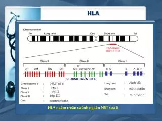
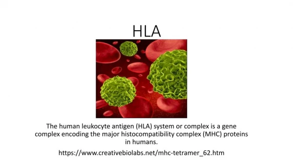
The HLA-A (hla a antibody) gene is found on the short arm of chromosome 6 and codes for HLA-bigger, Au2019s -chain component. The HLA-A-variation chainu2019s is crucial to HLA function.

E N D
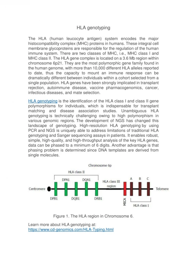
HLA A Structure — Boster Bio The HLA-A (hla a antibody) gene is found on the short arm of chromosome 6 and codes for HLA-bigger, A’s -chain component. The HLA-A-variation chain’s is crucial to HLA function. This variance encourages population genetic diversity. Because each HLA has a varied affinity for peptides with certain structures, having a larger number of HLAs allows for a greater diversity of antigens to be ‘presented’ on the cell surface, increasing the chances that a portion of the population will be resistant to a foreign invader. As a result, the chances of a single infection wiping out the whole human population are reduced. Each person can express up to two different kinds of HLA-A, one from each parent. Some people will inherit the same HLA-A (hla a antibody) from both parents, reducing their HLA variety; nevertheless, the vast majority of people will inherit two copies of HLA-A. All HLA groups follow the same pattern. To put it another way, each person can only express one or two of the 2432 HLA-A alleles. The HLA-A signal peptide is a group of hydrophobic amino acids found at the protein’s N-terminus that sends the protein to the endoplasmic reticulum, where the next seven domains are translated. The binding groove between the three domains retains a peptide for presentation to CD8+ t-cells. The trans-membrane region is the part of the phospholipid bilayer that surrounds the ER lumen that is embedded in the phospholipid bilayer. The HLA-A protein is a transmembrane protein with a single pass. In other words, the protein’s initial four domains are found inside the ER lumen, while the latter three domains are found outside the lumen, providing the protein with the appropriate orientation for function. The
protein’s last three domains form a tail of mainly -sheets that remains in the cell’s cytoplasm.