Download
1 / 30
300 likes | 318 Views
This lecture provides an anatomical review of the body's position and planes, as well as an in-depth exploration of the shoulder anatomy. It covers the movement of body parts, naming of muscles, and muscle terminology.

E N D
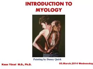
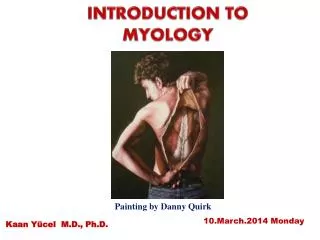
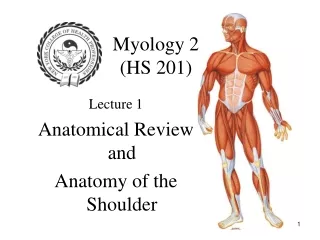
Myology 2 (HS 201) Lecture 1 Anatomical Review and Anatomy of the Shoulder
Anatomical Position Position In which the body is standing upright, the feet parallel, the arms hanging by the sides, and the palms and face directed forward.
PLANES OF THE ANATOMICAL POSITION A. Median (Mid-Sagittal): divides the body into symmetrical right and left halves. B. Sagittal: any plane parallel to the median plane C. Coronal: any vertical plane perpendicular to the median plane. It divides the body into anterior and posterior parts. D. Transverse: divides the body into superior and inferior parts E. Oblique: any plane on an angle
Movement of Body Parts • Flexion: movement in a sagittal plane which takes a part of the body forward from the anatomical position, causing a decrease in the angle of a joint. • Extension: movement in a sagittal plane which takes a part of the body backward from the anatomical position causing an increase in the angle of a joint. • Abduction: a movement in the frontal plane which takes a part of the body away from the median plane. ** For the fingers and toes the reference points used is the axis of the hand (middle finger) or foot (second toe). • Adduction: a movement in the frontal plane which takes a part of the body toward the median plane.
Movement of Body Parts • Horizontal Abduction: movement in a transverse plane with the arm or leg beginning 90 degrees from the trunk, taking the arm or leg away from the midline. • Horizontal Adduction: movement in a transverse plane with the arm or leg beginning 90 degrees from the trunk, taking the arm or leg toward the midline. • Lateral (External) rotation: movement of an extremity in a transverse plane which takes a body part outward. • Medial (Internal) rotation: movement of an extremity in a transverse plane which takes a part of the body inward.
Movement of Body Parts • Circumduction: combo of flexion-extension and abduction-adduction in succession • Supination: of the forearm, the palm faces forward • Pronation: of the forearm, the palm faces backward • Inversion: of the foot, soles facing inward • Eversion: of the foot, soles facing outward • Plantarflexion: of the foot, toes pointing down • Dorsiflexion: of the foot, toes point upward
Movement of Body Parts • Lateral flexion: applies to the head, neck or trunk. Have • movement in the frontal plane away from the median plane. • Rotation: applies to the head, neck or trunk. Movement in a • transverse plane where the body part turns either to the left or • right. • Protraction: drawing a structure forward • Retraction: drawing a structure backward
Other Anatomical Reference Terms a. Medial: closer to the median plane b. Lateral: further from the median plane c. Anterior: facing or located to the front d. Posterior: facing toward or located at the back e. Superior: facing toward or located at the top (closer to the head) f. Inferior: facing toward or located at the bottom (further from the head) g. Proximal: closer to the trunk or some major joint h. Distal: further from the trunk or some major joint i. Superficial: near the outside surface of the body, particular bone, or organ. j. Deep: inside the body, particular bone, or organ
NAMING OF MUSCLES 1. Direction of muscle fibers: a. Rectus: fibers run parallel to the midline (rectus abdominis) b. Transverse: fibers run perpendicular to the midline (transverse abdominis) c. Oblique: fibers run diagonally to the midline (external oblique) 2. Location: a. named for a nearby bone: (frontalis) frontal bone b. position relative to a bone: (tibialis anterior) tibia c. section of a muscle having more than one part (anterior deltoid)
NAMING OF MUSCLES (cont) 3. Size: a. large muscle (gluteus maximus) b. small muscle (gluteus minimus) c. long muscle (extensor digitorum longus) d. short muscle (peroneus brevis) 4. Number of origins: a. two origins: biceps femoris b. three origins: triceps brachii c. four origins: pronator quadratus
NAMING OF MUSCLES(cont) 5. Shape: a. triangular: deltoid b. trapezoid: trapezius c. saw-toothed: serratus d. rhomboid or diamond shaped: rhomboids 6. Origin and Insertion: a. sternum, clavicle, and mastoid process: SCM
NAMING OF MUSCLES (cont) 7. Action: a. decreases an angle of a joint: flexor digitorum b. increases an angle of a joint: extensor carpi ulnaris c. moves bone away from midline: abductor pollicis brevis d. moves bone closer to midline: adductor longus e. upward movement: levator scapulae f. downward movement: depressor labii g. turns palm up or anteriorly: supinator h. turns palm down or posteriorly: pronator i. decreases size of an opening: external anal sphincter j. makes a body part more rigid: tensor fascia latae k. moves a bone around its longitudinal axis: rotator
MUSCLE TERMINOLOGY 1. Belly: main portion of muscle which contains contractile fibers 2. Tendon: connective tissues which attaches muscle to the bone (they resist pulling) 3. Aponeurosis: tendon that fans out and attaches to a broad area. 4. Insertion: movable end of a muscle attached to a bone that is being moved 5. Origin: fixed end of a muscle which does not move 6. Fascia: connective tissue that holds fibers of muscles in place (also hold organs)
Depressions and openings: Fissure: narrow opening between adjacent parts of bones for nerves and vessels Foramen: hole, opening Fossa: shallow depression Sulcus: groove Meatus: tubelike passageway Processes that form joints Condyle: large rounded prominence Facet: smooth flat surface Head: rounded articular projection Processes for tendon and ligament attachment: Crest: prominent border or ridge Epicondyle: prominence above a condyle Linea: line, less prominent than a crest Trochanter: large projection of bone found only on the femur Tubercle: small rounded process Tuberosity: large, rounded, usually roughened process Bone Surface Markings
MUSCLE ACTIONS 1. Agonist: main muscle that moves a joint 2. Antagonist: opposes action of the agonist, moves the muscle back to place, there is balance between the agonist and antagonist 3. Synergist: muscles acting in a coordinated fashion to provide a movement. Examples: levator scapulae and trapezius cause elevation of scapula, gastrocnemius, soleus, and plantaris cause us to step on toes 4. Fixator: muscle used to stabilize bones. Holds bone in place so other muscles can act. 5. Neutralizer: Two muscles that cancel each other out in one action but work synergistically to perform another action.
Shoulder Girdle • Shoulder girdle consists of the scapula and the clavicle • Function of the shoulder girdle is to attach the upper extremity to the trunk • The scapula and clavicle are united at the acromioclavicular joint (a gliding joint)
Scapula • Triangular bone, located on posterior thorax between levels of 2nd and 7th ribs • Anatomy: • Scapular spine: sharp ridge of bone, running diagonally across the posterior surface of the scapula. • Body: main triangular portion of the scapula • Acromion: flattened lateral end of the scapular spine (high point of the shoulder). • Acromion process articulates with the lateral end of the clavicle • Glenoid cavity (fossa): depression on the lateral end of the scapula for articulation with the head of the humerus • Medial (vertebral) border: thin edge of the scapular body near vertebral column • Lateral (axillary) border: thicker edge of scapular body on the lateral side
Scapula • Anatomy (cont): • Inferior angle: angle formed by the joining of the medial and lateral borders • Superior border: superior edge of scapula • Superior angle: angle formed by the joining of the superior border and medial border. • Scapular notch: indentation in superior border for suprascapular nerves • Coracoid process: hooked projection of bone from the anterior surface of the scapula. Functions as a point of muscle attachment. • 3 fossae: • Supraspinous fossa: fossa above the scapular spine • Infraspinous fossa: fossa below scapular spine • Subscapular fossa: fossa on the anterior surface of the scapula
Clavicle • Long slender S-shaped bone known as the collarbone • Medial 1/3 of clavicle is convex anteriorly, Lateral 1/3 is concave • Junction of the two curves is weakest point and subject to fractures. • Anatomy: • Sternal end: medial rounded end of clavicle. • Articulates with sternum forming the sternoclavicular joint. • Acromial end: broad, flat, lateral end of clavicle • Articulates with acromion process of scapula forming the acromioclavicular joint (A-C joint). • Conoid tubercle: on inferior lateral end of clavicle for ligamentous attachment • Costal tuberosity: on inferior medial end of clavicle also for ligamentous attachment.
Proximal Humerus • Anatomy: • Head: rounded surface articulating with glenoid fossa • Anatomical neck: groove distal to the head of the humerus • Greater tubercle: lateral projection distal to the neck • Lesser tubercle: anterior projection of bone • Intertubercular sulcus (Bicipital groove): groove between the greater and lesser tubercle. Receives the long head of the biceps muscle. • Surgical neck: constricted portion distal to the two tubercles • Named so because a common site of humeral fracture • Shaft: diaphysis of the arm bone • Deltoid tuberosity: roughened V-shaped area of bone on the lateral portion of the shaft
Articular Anatomy of the Shoulder 1. Sternoclavicular Joint: gliding joint where head of clavicle fits into notch on sternum. 2. Acromioclavicular Joint: gliding joint where clavicle articulates with acromion process of scapula. Shoulder separation occurs at the AC joint 3. Glenohumeral joint: ball and socket joint between glenoid fossa of scapula and head of the humerus. Shoulder dislocation takes place at the glenohumeral joint. 4. Scapulothoracic joint: where anterior scapula articulates with the ribs
Soft Tissue Anatomy of the Shoulder 1. Rotator Cuff: attachment of rotator cuff muscles around the shoulder joint capsule. It is thick and strong. 2. Biceps Tendon: attaches biceps muscle to scapula 3. Bursa: fluid filled sacs which lubricates tendons as they move across each other: a. subscapular bursa b. subdeltoid bursa c. subacromial bursa d. subcoracoid bursa
Soft Tissue Anatomy of the Shoulder (Cont) Ligaments: 1. Coracohumeral ligament: is a strong, broad ligament that strengthens the superior part of the articular capsule. Extends from the coracoid process of the scapula to the greater tubercle of the humerus. 2. Acromioclavicular ligament: lateral clavicle to superior surface of acromion 3. Coracoacromial ligament: a scapular ligament. From coracoid to acromion; strengthens superior articular capsule
Soft Tissue Anatomy of the Shoulder (Cont) Ligaments (cont): 4. Coracoclavicular ligament: strong ligament divided into two ligaments (conoid and trapezoid) They anchor the lateral part of the clavicle to the coracoid process. a. Conoid: passes from the coracoid process to the clavicle. Resists forward movement of the scapula without corresponding movements of the clavicle. b. Trapezoid: resists backward movement of the scapula.
Soft Tissue Anatomy of the Shoulder (Cont) Ligaments (cont): 5. Costoclavicular ligament: from first costal cartilage to medial clavicle. Limits elevation of the clavicle. 6. Sternoclavicular ligament: from sternum to clavicle. Reinforces articular capsule of sternoclavicular joint. 7. Interclavicular ligament: extends across jugular notch. Stabilizes the sternoclavicular joint.
Soft Tissue Anatomy of the Shoulder (Cont) 8. Articular capsule: is a thin, loose sac that completely envelopes the shoulder joint. It extends from the glenoid cavity to the anatomical neck of the humerus. The inferior part of the capsule is its weakest area. 9. Glenoid Labrum: a rim of fibrocartilage around the edge of the glenoid cavity. It functions to deepen and enlarge the glenoid cavity