Download
1 / 36
520 likes | 1.05k Views
Microcytic Anemias. Sex & Gender Difference Slides. When in presentation mode, click this button to advance to each sex and gender difference slide. Overview. Microcytic Anemias. Anemia is defined as: hemoglobin <12 g/dL in females and <13 g/dL in males 1 Anemias based on morphology: 2

E N D
Sex & Gender Difference Slides When in presentation mode, click this button to advance to each sex and gender difference slide.
Overview Microcytic Anemias
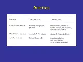
Anemia is defined as: • hemoglobin <12 g/dL in females and <13 g/dL in males1 • Anemias based on morphology:2 • Microcytic- MCV <80 fL • Normocytic- MCV 80-100 fL • Macrocytic- MCV >100 fL • Anemia is more prevalent in females3 Anemia Overview
Most Common Causes of Anemias in Females Worldwide:1 • Iron deficiency anemia due to: menstrual blood loss and childbirth, uterine bleeding disorders • Hemoglobinopathies • GI losses • Gynecological conditions • Other nutritional deficiencies • Anemia second to chronic disease • Aplastic anemia • Anemia secondary to infiltrative disorder Anemias in Females
Most Common Causes of Anemias in Males Worldwide:1 • Iron deficiency anemia • Hemoglobinopathies • GI losses • Tropical diseases Anemias in Males bleeding from duodenal ulcer
Caused by Decreased Hemoglobin Production1,2 • Types:1,2 • Iron deficiency • Thalassemias • Hemoglobinopathies • Anemia of chronic disease Thalassemia peripheral blood smear Microcytic Anemia
Iron Deficiency Anemia
Affects >20% of reproductive aged females2 • Affects <1% of males in the same age group4 • Global prevalence 38% in pregnant women2 • 40% prevalence in preschool aged children3 Epidemiology
Adolescent/adult females of childbearing age every 5-10 years1 • No screening needed for males or post-menopausal females1,2 • First prenatal visit1,2 Screening
Asymptomatic • Pallor • Fatigue • Most common presentation in females • Koilonychia • Pica Koilonychia Symptoms Pallor
Occult blood loss • Insufficient oral intake of iron • Poor gastrointestinal absorption of iron • Pregnancy and delivery Causes
Decreased in premenopausal and menstruating females1,2 • Postmenopausal females and males exhibit similar levels1,2 Hepcidin
Diagnosis Anemia
Treatment Anemia
Oral iron supplementation1,2 • Increase dietary iron intake1,2 Treatment
Impaired cognitive function1 • Perinatal complications1 • Increased falls in the elderly1 • Strong predictor of GI cancer in males and postmenopausal females2 Colon cancer Complications
Associated with increased maternal and neonatal morbidity and mortality1 • Increased risk of:1 • Prematurity • Low birth weight • Peripartum blood loss Pregnancy and Anemia
Thalassemias Anemia
Absent or Defective α chain Production1,2 α-Thalassemia Pathogenesis
Absent or Defective β chain Production1,2 β-Thalassemia Pathogenesis
Diagnosis Anemia
Hemoglobin Electrophoresis • Peripheral Smear Showing:3 • Microcytes (M) • Target cells (T) • Poikilocytes (P) β-Thalassemia Diagnosis
Hepatosplenomegaly • Growth Impairment • Bone abnormalities • Hypogonadotropic hypogonadism • Hypercoagulability • Pulmonary Hypertension Expansion of the medullary space and new frontal bone formation Thalassemia Complications
Treatment Anemia
Transfusion • Splenectomy • Iron Chelation • Fetal Hemoglobin Inducers • Stem Cell Transplant Thalassemia Treatment
Reproduction Anemia
α-thalassemia retardation (X-linked):1,2,3 • Occurs only in males • Severe mental retardation • Developmental abnormalities • Genital abnormalities • Classic facies ATR-X
Caused by iron overload secondary to transfusions and gut absorption1,2 • Iron deposition in the pituitary1,2 • Iron chelation therapy1 • Ovulation induction1 Hypogonadotropic Hypogonadism and Fertility
If undiagnosed in mom:1,2,3 • CBC in prenatal labs • Hemoglobin electrophoresis If undiagnosed in both parents:1,3 • Prenatal ultrasound If mutations have been identified in both parents:2,3 • Preimplantation genetic diagnosis with IVF • Chorionic villus sampling or amniocentesis Hydrops fetalis Prenatal Diagnosis
Cardiac impairment • Anticoagulation • Vertical transmission of viruses • Alloimmunization • Fetal Loss Thalassemia and Pregnancy