Download
1 / 21
210 likes | 234 Views
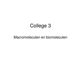
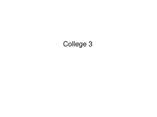
College 3. Fig.2.1.26. Hückel molecular orbitals for 1.3-butadiene. The orbitals are viewed perpendicular to the plane of the molecule. The carbon nuclei are represented by dots, and the nodal planes for the MO’s are represented by the dashed lines.

E N D
Fig.2.1.26. Hückel molecular orbitals for 1.3-butadiene. The orbitals are viewed perpendicular to the plane of the molecule. The carbon nuclei are represented by dots, and the nodal planes for the MO’s are represented by the dashed lines. With each carbon atom contributing one electron the 1p and 2p orbitals are filled. Huckel voor 1,3 butadiene C atomen: 3 sp2 hybidide orbitalen h1, h2, h3 CH2=CHCH=CH2 2 e- in 1s 3 e- in h1, h2, h3 1 e- over voor π orbitaal Voor alle 4 C atomen, dus 4e
Vergelijk ethyleen met butadieen Als butadieen = 2 x ethyleen Dan bindingsenergie is 4α+4β, Maar door delocalisatie: 2(α+1.618 β) + 2(α+0.618 β)= 4 α+4.472 β Verschil: 0.472 β
In plaats van Nog groter: benzeen
The answer: only with A & T and with C & G are there opportunities to establish hydrogen bonds (shown here as dotted lines) between them (two between A & T; three between C & G). These relationships are often called the rules of Watson-Crick base pairing, named after the two scientists who discovered their structural basis. The rules of base pairing tell us that if we can "read" the sequence of nucleotides on one strand of DNA, we can immediately deduce the complementary sequence on the other strand. DNA base pairing • A with T: adenine (A) always pairs with thymine (T) • C with G: cytosine (C) always pairs guanine (G) • This is consistent with there not being enough space (20 Å) for two purines to fit within the helix and too much space for two pyrimidines to get close enough to each other to form hydrogen bonds between them. • But why not A with C and G with T?
Non-Covalent Interactions that determine 3-D structures of proteins, membranes… When two neutral molecules come close to one another (even when they are non-polar)….. Attraction ~ kBT Fig.2.2.1 The energy of interaction of two N2-molecules as a function of distance according to the Lennard-Jones potential. Note that the ordinate is in kelvins; the interaction energy has been divided by the Boltzmann constant.
Charge-Charge and dipole-dipole interactions Molecules with charges, come close → interaction energy is given by Coulombs law, 1/r ‘long range interaction’ Potential energy (at 1 by 2) In water Coulomb interactions are reduced by a factor, which is the dielectric constant of water. (water molecules have a large permanent dipole which effectively screens the charges). In a protein environment, ion pairs with opposite charges may contribute significantly to the stabilization of a biomolecular structure. Of course on an atomic scale the concept of the dielectric constant becomes meaningless and one would have to calculate explicitly all the interactions between the charges.
Charge-Charge and dipole-dipole interactions Molecules with no net charges, also attract each other, when they have an asymmetric charge distribution, resulting in a dipole μ. μ1 μ2 interaction affects potential energy Fig 2.2.2 (a) Attractive dipole-dipole interaction between two molecules. (b) repulsive counterpart of (a). If molecules are randomly oriented the average interaction energy is zero. But: attractive orientations are energetically more favorable
Charge-Charge and dipole-dipole interactions includes a weighting factor in the averaging that is equal to the probability that the molecules (dipoles) will adapt a certain orientation. This probability is given by the Boltzmann factor , with E interpreted as the potential energy of interaction of the two dipoles in that orientation. This implies that for every orientation f we have: with Als V<< KT, dan kunnen we dit ontwikkelen leidende tot turns out to be 2/3
Charge-Charge and dipole-dipole interactions Belangrijk: R6, 1/T (greater thermal motion overcomes the mutual orientating effects of the dipoles at higher temperatures) Averaging only for ‘freely moving’ solvents In proteins a typical electrostatic calculation puts partial charges on all of the atoms (orgroups of atoms) and simply solves the Poisson equation numerically
Van der Waals krachtennon-charged, non-ionic molecules • permanent dipole–permanent dipole forces √ • permanent dipole–induced dipole forces • instantaneous induced dipole-induced dipole
Van der Waals krachtennon-charged, non-ionic molecules • permanent dipole–induced dipole forces If molecule A has a dipole moment this creates an electric field which polarizes the charge distribution on molecule B → induced dipole moment μB= α = polarizability of molecule B. μB is always oriented in the direction of the electric field created by μAand consequently the interaction is always attractive. A calculation of this interaction yields:
Van der Waals krachtennon-charged, non-ionic molecules How do non-polar molecules form condensed phase??? For example, hydrogen or argon condensate to a liquid at low temperatures and benzene is a liquid at normal temperatures • permanent dipole–permanent dipole forces √ • permanent dipole–induced dipole forces √ • instantaneous induced dipole-induced dipole
Van der Waals krachtennon-charged, non-ionic molecules • permanent dipole–permanent dipole forces √ • permanent dipole–induced dipole forces √ • instantaneous induced dipole-induced dipole Coupling of instantaneous fluctuations in the charge distribution on one molecule that gives it a dipole briefly; that dipole may induce another dipole in a neighbouring molecule, and these two dipoles will interact favorably. Also in proteins Van der Waals forces and specifically the dispersion force, plays a major role in the formation of their stable and active conformation, in particular in stabilizing the hydrophobic core.
Fig. 2.2.3 Typical form of the Lennard Jones potential. The distance at which the minimum occurs would be two times the van der Waals radius of the molecules involved. The intermolecular potential energy At small distances the molecules repel strongly, (we try to push the electrons of one molecule into the non-bonding orbitals of the other, the bonding orbitals are occupied by electron pairs), ~R-n
The hydrogen bond Fig. 2.2.5.A hydrogen bond between two water molecules. The strength of the interaction is maximal when the O-H covalent bond points directly along a lone-pair electron cloud of the oxygen atom to which its hydrogen bonded. In het algemeen: D—H · · · ·A The large electronegativity difference between H and O confers a 33% ionic character on the OH-bond as reflected by water’s dipole of 1.85 debye units. → highly polar molecule The electrostatic interactions between the dipoles of two water molecules tend to orient them such that the O-H bond on one molecule points towards a lone pair electron cloud on the oxygen atom of the other water molecule H-bond distances 0.5 Å shorter than van der Waals distance! Energy of the hydrogen bond, about 20kJmol-1 in H2O, is small compared to covalent bond energies (for instance 460 kJmol-1 for an O-H covalent bond)
The hydrophobic interaction Because these cage structures are more ordered than the surrounding water, their formation increases the free energy. This free energy cost is minimized, however, if the hydrophobic (or hydrophobic parts of amphipathic molecules) cluster together so that the smallest number of water molecules is affected.