Download
1 / 19
190 likes | 433 Views
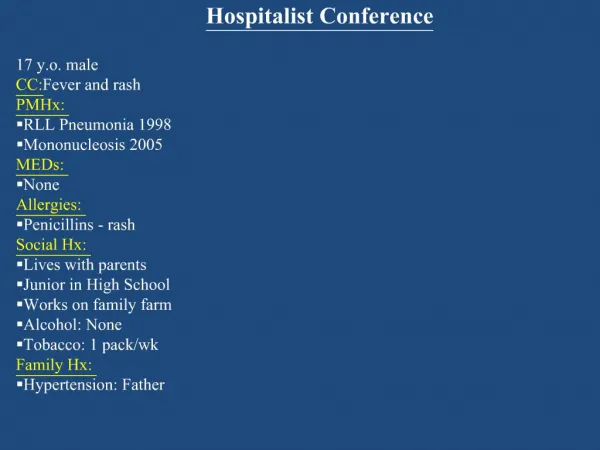
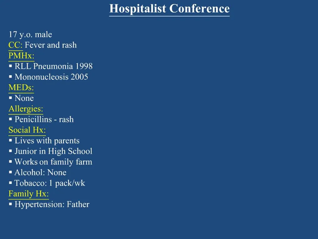
17 y.o. maleCC: Fever and rashPMHx: RLL Pneumonia 1998 Mononucleosis 2005MEDs: NoneAllergies: Penicillins - rashSocial Hx: Lives with parents Junior in High School Works on family farm Alcohol: None Tobacco: 1 pack/wkFamily Hx: Hypertension: Father Colon Cancer: PGF. Case Conference.

E N D
8. Leptospirosis
9. History A zoonosis caused by the spirochete leptospira interrogans
1883: First recognized as an occupational disease of sewer workers
1886: Weil�s disease
Named after Adolph Weil who described the disease as: �an acute infectious disease with enlargement of spleen, jaundice, and nephritis�
This is most severe form of leptospirosis
1907: Stimpson, first isolate
10. Epidemiology Worldwide distribution
Most cases occur in tropics
Thailand: 30-fold increased in cases from 1995-2000
Hypothesis: increased rat population and seasonal flooding
In US, most cases are in southern and Pacific coastal states
Hawaii has most cases of any state in US
Outbreaks can occur
12% of athletes participating in Illinois triathlon after exposure to lake water in swimming phase
Areas with high rat population and seasonal flooding have the highest incidence
11. At Risk Populations Occupational Exposure:
Farmers, veterinarians, sewer workers, rice field workers
Recreational Activities:
Fresh water swimming, canoeing, kayaking
Household Exposures:
Domesticated livestock, infestation by infected rodents
12. Pathogenesis Humans become infected after exposure to environmental sources:
Animal urine (wild and domestic mammals especially rodents, cattle, swine, dogs, horses, sheep, and goats)
Contaminated soil or water
Infected animal tissue
Portals of entry:
Abraded skin
Mucous membranes
Conjunctiva
Incubation period 7-12 days
13. Clinical Course 90% of patients have mild symptoms while 5-10% have severe form with jaundice (Weil�s Disease)
Natural course has 2 distinct phases:
First Stage (Leptospiremic): Lasts 4-7 days
Non-specific flu-like symptoms
Fevers, chills, sore throat, headaches, myalgias, rash
Second Stage (Immune or Leptospiruric): Lasts up to 30 days
Circulating antibodies may be detected
Organism may be isolated from urine
Meningeal symptoms in 50% of patients
Viral etiology may be suspected
14. Exam findings During First Stage:
Fevers, pharyngeal injection, lymphadenopathy
Conjunctival suffusion:
Conjunctival redness due to increased blood flow
During Second Stage:
Adenopathy, rash, fever
Jaundice, splenomegaly, abdominal tenderness
15. Advanced Disease � Weil�s Syndrome Severe form of leptospirosis characterized by profound jaundice, renal dysfunction, hepatic necrosis, and hemorrhagic diathesis
Criteria for diagnosis are not well defined
Complications include:
Renal failure, uveitis, hemorrhage, ARDS, myocarditis, rhabdomyolysis, liver failure
Mortality rate of 5-10%
Some studies suggest case fatality rates of 20-40%
16. Laboratory Findings Thrombocytopenia
Leukocytosis with left shift
Elevations of transaminases (<200) in 40% of patients
Elevated CK in up to 50% of patients
UA with proteinuria
CSF may show a neutrophilic or lymphocytic pleocytosis with normal protein and glucose
17. CDC Diagnostic Criteria
18. Diagnosis Culture:
Blood
Positive in 1st 10 days of illness
Isolation successful in only 50% of cases
CSF
Positive in 1st 10 days of illness
Urine
Becomes positive in 2nd week of illness
May remain positive for up to 30 days after resolution of symptoms
19. Serology:
Microscopic agglutination test (MAT), macroscopic agglutination test, indirect hemagglutination, and ELISA
Gold standard is MAT, but is not widely available
Most common tests used in clinical practice:
Microplate IgM ELISA
IgM dot-ELISA dipstick
If one of these is positive, sera for MAT can be sent to CDC
PCR is being explored and showing some promise in diagnosis, but is not yet widely available Diagnosis
20. Treatment Antibiotic treatment for one week
Doxycycline 100 mg IV or po q 12 hrs
Ampicillin 500 - 1000 mg IV q 6 hrs
Penicillin G 3-4 million units IV q 4 hrs
Penicillin G 1.5 million units IV q 6 hrs
Ceftriaxone 1 gram IV qd