Insight into Visual Pigments and Biochemistry of Human Vision
Explore the fascinating world of visual pigments and biochemistry related to human vision, including mechanisms of activation, amplification, and termination. Learn about the basics of how visual transduction works in dim vs. bright light, the metabolism of vitamin A, and the intricacies of color vision. This comprehensive guide references key sources in biochemistry and molecular biology to provide a deeper understanding of the components and players involved in the visual process.

Insight into Visual Pigments and Biochemistry of Human Vision
E N D
Presentation Transcript
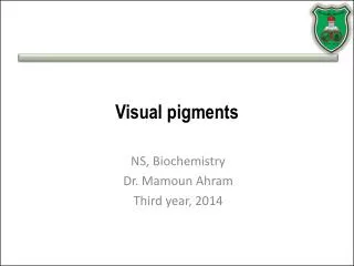
Visual pigments NS, Biochemistry Dr. Mamoun Ahram Third year, 2014
References • Photoreceptors and visual pigments • Webvision: The Organization of the Retina and Visual System (http://www.ncbi.nlm.nih.gov/books/NBK11522/#A127) • Molecular Biology of the Cell. (http://www.ncbi.nlm.nih.gov/books/NBK26912/#A2826) • Biochemistry (http://www.ncbi.nlm.nih.gov/books/NBK22541/#A4618) • Vitamin A and Carotenoids • Lippincott Williams & Wilkins, p.381-383
Lecture outline • Visual transduction (dim vs. bright light) • Components (cells and molecules) • Mechanisms of activation, amplification, and termination • Color blindness • Metabolism of vitamin A
Rods and cones Dim Light (1 photon) Bright Light 7 million 120 million
The dark current Na+ and a lesser amount of Ca2+ enter through cyclic nucleotide-gated channels in the outer segment membrane K+ is released through voltage-gated channels in the inner segment. Rod cells depolarize. The neurotransmitter glutamate is released continuously. Channels in the outer segment membrane close, the rod hyperpolarizes Glutamate release decreases.
The players • Rhodopsin • Transducin • Phosphodiesterase • Na+-gated channels • Regulatory proteins
Rhodopsin chromophore
11-cis-retinal 10-13 sec
Rhodopsin intermediates • Rearrangements in the surrounding opsin convert it into the active R* state. • The chromophore converts the energy of a photon into a conformational change in protein structure.
Transducin G proteins are heterotrimeric, consisting of , , and subunits. In its inactive state, transducin’s subunit has a GDP bound to it.
Transducin GTP R* binds transducin and allows the dissociation of GDP, association of GTP, and release of the subunit.
Activation of phosphodiesterase • PDE is a heterotetramer that consists of a dimer of two catalytic subunits, and subunits, each with an active site inhibited by a PDE subunit. • The activated transducin subunit-GTP binds to PDE and relieves the inhibition on a catalytic subunit.
cGMP-gated channels Ca2+ • When activated, PDE hydrolyzes cGMP to 5’-GMP • The cGMP concentration inside the rod decreases • Cyclic nucleotide-gated ion channels respond by closing
Animation movie http://www.ncbi.nlm.nih.gov/books/bookres.fcgi/webvision/photomv3-movie1.mov
Facilitation of transduction 2-dimensional surface low in cholesterol and have a high content of unsaturated fatty acids
Cooperativity of binding The binding of one cGMP enhances additional binding and channel opening (n = about 3) • Overall, a single photon closes about 200 channels and thereby prevents the entry of about a million Na+ ions into the rod.
Mechanism IArrestin binding • Rhodopsin kinase (GRK1) phosphorylates the C-terminus of R*. • Phosphorylation of R* decreases transducin activation and facilitates binding to arrestin, which completely quenches its activity, and release of the all trans-retinal regenerating rhodopsin.
Mechanism IIArrestin/transducin distribution • In dark, the outer segment contains high levels of transducin and low levels of arrestin. • In light, it is the opposite.
Mechanism IIIGTPase activity of G protein • Transducin has an intrinsic GTPase activity that hydrolyzes GTP to GDP. • Upon hydrolysis of GTP to GDP, transducin subunit releases the PDE subunit that re-inhibits the catalytic subunit. • Transducin -GDP eventually combines with transducin
A role for calcium ions When the channels close, Ca2+ ceases to enter, but extrusion through the exchanger continues, so [Ca2+]int falls.
500 nM Mechanism VGuanylate cyclase 50 nM • In the dark, guanylate cyclase-associated proteins (GCAPs) bind Ca2+ and inhibit cyclase activity. • A decrease in [Ca2+]int causes Ca2+ to dissociate from GCAPs, allowing them to dimerize. • Dimerization of GCAPs leads to full activation of guanylate cyclase subunits, and an increase in the rate of cGMP synthesis
Mechanism VICa-calmodulin • In the dark, Ca2+-Calmodulin (CaM) binds the channel and reduces its affinity for cGMP. • During visual transduction, the decrease in [Ca2+]int causes CaM to be released, increasing the channel’s affinity for cGMP so that during recovery, the channel reopens at lower levels of cGMP
How different are they? • Cone opsins have similar structures as rhodopsin, but with different amino acid residues surrounding the bound 11-cis retinal; thus they cause the chromophore’s absorption to different wavelengths. • Each of the cone photoreceptors vs rhodopsin 40% identical. • The blue photoreceptor vs green and red photoreceptors = 40% identical. • The green vs. red photoreceptors > 95% identical.
Three important aa residues A hydroxyl group has been added to each amino acid in the red pigment causing a max shift of about 10 nm to longer wavelengths (lower energy).
Rods vs. cones • Light absorption, number, structure, photoreceptors, chromophores, image sharpness, sensitivity
Chromosomal locations • The "blue" opsin gene: chromosome 7 • The "red" and "green" opsin genes: X chromosome • The X chromosome normally carries a cluster of from 2 to 9 opsin genes. • Multiple copies of these genes are fine.
Red-green homologous recombination • Between transcribed regions of the gene (inter-genic) • Within transcribed regions of the gene (intra-genic)
Examples Red blindness Green blindness