Download
1 / 58
730 likes | 1.43k Views
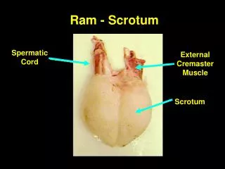
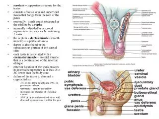
scrotum = supportive structure for the testes consists of loose skin and superficial fascia that hangs from the root of the penis externally- single pouch separated at the midline by a raphe internally – divided by a scrotal septum into two sacs each containing 1 testis

E N D
scrotum = supportive structure for the testes • consists of loose skin and superficial fascia that hangs from the root of the penis • externally- single pouch separated at the midline by a raphe • internally – divided by a scrotal septum into two sacs each containing 1 testis • the septum = dartos muscle (smooth muscle) + superficial fascia • dartos is also found in the subcutaneous portion of the scrotal skin • each testis is associated with a cremaster muscle – skeletal muscle that is a continuation of the internal oblique • exterior location of the testis ensures its internal temperature is at least 2 to 3C lower than the body core • failure of the testes to descend = cryptochidism • 3% of full-term infants and 30% or premature infants • untreated – results in sterility • increases the chances of testicular cancer • 80% of those undescended testes will descend spontaneously within the year
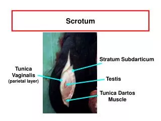
-testis: develop internally near the kidneys and descend through the inguinal canal during the latter half of the seventh month gestation -covered by several protection membranes 1. tunica vaginalis – serous membrane derived from the peritoneum, forms during the descent of the testes -injury to the testes can cause an accumulation of fluid within the membrane = hydrocele -allows for easier movement of the testes within the scrotum 2. tunical albuginea – internal to the TV -extends inward to divide the testes into lobules (200-300) -each lobule contains 1 to 3 coiled seminiferous tubules for sperm production -lined with epithelium that produce sperm (spermatogenic cells)
embedded among the spermatogenic cells of the seminiferous tubules – Sertoli cells • sustenacular cells • extend from the basement membrane of a seminiferous tubule to the lumen • during the Maturation phase of spermiogenesis - Sertoli cells consume the unneeded portions of the spermatazoa. • once fully differentiated, the Sertoli cell is unable to proliferate • adjacent cells are joined together by tight junctions – blood-testis barrier • this barrier prevents an immune response against the spermatogenic cells’ surface antigens which are recognized as they develop as being foreign • this creates a privileged immune environment • also secrete numerous cytokines and growth factors that mediate spermatogenesis • anti-Müllerian hormone (AMH) - secreted during the early stages of fetal life. • inhibin and activins - secreted after puberty, and work together to regulate FSH secretion • androgen binding protein - faciliate spermatogenesis and sperm maturation • glial cell line-derived neurotrophic factor (GDNF) - has been demonstrated to function in promoting undifferentiating spermatogonia - ensures stem cell self-renewal • the Ets related molecule (ERM transcription factor) - needed for maintenance of the spermatogonial stem cell in the adult testis. • transferrin • between adjacent seminiferous tubules are the interstitial cells or Leydig cells • for the production of testosterone (androgen) • can be a site for the development of testicular cancer - along with Sertoli cells • androgen = hormone for the development of masculine characteristics • Sertoli-Leydig tumor – type of ovarian tumor • Arrhenoblastomas – Sertoli-Leydig cells within the ovary secrete androgens leading to virilization of the female phenotype
Medical application • Sertoli cells & the immune system • presence of a privileged immune environment within the testes • Sertoli cells can be transplanted into an ectopic site – recreates this immune environment • other cells and tissues co-transplanted into these ectopic sites are protected from the immune system • transplantation of canine pancreatic islets – production of insulin • also secrete growth factors that improve the engraftment, vascularization and survival of the co-transplanted tissues
Spermatogenesis • sperm development – from sperm stem cells called spermatogonium • these spermatogonium develop in the embryonic testes from primordial germ cells that arise from the yolk sac • the spermatogonium remain dormant in the testes until puberty • the maturing sperm can be found toward the lumen of the seminiferous tubule • most mature = sperm cells or spermatozoa • takes 60-75 days to complete • 1. dissociation of some spermatogonium from the basemement membrane of the ST • 2. differentiation of the dissociating spermatogonium into primary spermatocytes (2n) • 3. replication of DNA within the spermatocyte and the onset of meiosis • 4. formation of secondary spermatocytes (n) – however despite having 23 chromosomes, these chromosomes are still comprise of two chromatids • 5. completion of meiosis and formation of spermatids (n) – 23 chromosomes each made up of one chromatid • 6. spermiogenesis – development of spermatids into a sperm cell • spherical spermatids transform into elongated sperm containing an acrosome and bearing a flagellum • as the spermatogenic cells form through meiosis they fail to undergho complete cytokinesis • cells remain in contact throughout meiosis via cytoplasmic bridges • accounts for the synchronization observed in the production of sperm in any given area of the seminiferous tubule
Sperm • 300 million made each day • 60 um long • major parts • 1. head: contains the nucleus with 23 highly condensed chromosomes (one chromatid) • 2. acrosome: covers the anterior 2/3 of the head • contains digestive enzymes to dissolve the protective barriers of the egg (hyaluronidase and proteases) • 3. tail or flagellum • neck - constricted region just behind the head • contains centrioles for the production of the microtubules for the tail • middle piece – contains mitochondria arranged in a spiral • principal piece – longest portion of the tail • end piece – terminal portion of the tail
-release of gonadotropic releasing hormone (GnRH) from the neurosecretory cells of • the hypothalamus which stimulates the gonadotrophs of the anterior pituitary gland • -anterior pituitary releases gonadotropins (FSH and LH) • Follicle stimulating hormone - stimulates spermatogenesis • -synergistic action by FSH and testosterone on the Sertoli cells – secrete androgen-binding protein into the lumen of the seminiferous tubule • -ABP binds to testosterone and keeps the concentration of this androgen high within the ST • -testosterone stimulates the final stages of spermatogenesis • -FSH release is inhibited by the release of inhibin by the Sertoli cells • 2. Leutinizing hormone - stimulates male hormone production by the Leydig cells • -testosterone synthesized from cholesterol in the testes • -suppresses GnRH synthesis by negative feedback • -in some targets (e.g. prostate), testosterone is converted into dihydrotestosterone (DHT)
Sertoli-Leydig cell interaction: Paracrine regulation • Sertoli cells exert a paracrine control of the two main testicular functions, androgen secretion and spermatogenesis • co-culture of Sertoli cells with Leydig cells • increases testosterone production by Leydig cells • causes Leydig cells to produce an increase in FSH receptors • coculture of rat or pig Sertoli cells with rat germ cells • induces an increase in the RNA and DNA biosynthetic activities of germ cells – increases spermatogenesis • most of the stimulatory effects of Sertoli cells seemed to be mediated by diffusible factors like FSH (paracrine), secreted by Sertoli cells, but full • but stimulation of germ cells seems to require cell-cell contact with Sertoli cells
Testosterone • testosterone and DHT both bind to same receptors • receptors are found within the nuclei of the target cells • targets – bone, muscle • effects • 1. prenatal development • stimulates the male pattern of the reproductive system • gonads develop during the 5th week of gestation from two sets of ducts: 1) Wolffian ducts (males) and 2) Mullerian ducts (females) • therefore the embryo has the potential to develop into either sex • BUT “maleness” determined by a gene called SRY – sex determining region of the Y chromosome • SRY protein expression induces differentiation of Sertoli cells • Sertoli cells secrete Mullerian-inhibiting Substance – apoptosis within the Mullerian ducts which inhibits the development of female structures -in response to hCG – Leydig cells begin to synthesize testosterone -testosterone stimulates development of the epididymus, vas deferens, ejaculatory duct and seminal vesicle • DHT stimulates development of external genitalia • development around the 8th week • from the genital tubercle (both males and females) – comprised of a urethral groove and two labiosacral swellings • elongation of part of the genital tubercle into the penis • labiosacral swellings - scrotum • testosterone is converted in the brain to estrogens – development of certain brain regions in males • 2. development of male sexual characteristics • 3. development of sexual function • male sexual behavior • spermatogenesis • libido in both males and females • females – androgen release by the adrenal cortex • 4. stimulation of anabolism • stimulate protein synthesis
Medical application: Anabolic steroids • a class of natural and synthetic steroid hormones • first discovered in the early 1930s • promote cell growth and division, protein sythesis (anabolism) • results in growth of several types of tissues, especially muscle and bone • increases bone remodelling and growth, increases bone marrow production of RBCs • increases size of clitoris or penis, increase vocal cord thickness, increases the libido, inhibits spermatogenesis • different anabolic androgenic steroids have varying combinations of androgenic and anabolic properties, and are often referred to in medical texts as AAS (anabolic/androgenic steroids) • for reversal of chronic wasting conditions including cancer and AIDS • stimulation of myogenesis • hypertrophy of both types of muscle fibers (I and II) • mechanism of this is not completely understood • increased synthesis of muscle proteins and/or decrease degradation of muscle proteins • also – increased commitment of muscle stem cells to the myogenic lineage and inhibiting their differentiation to the adipogenic • supraphysiological doses of testosterone in men promotes nitrogen density and increases fat free mass (skeletal muscle mass) while at the same time decreasing fat, particularly abdominal fat. • may also play an anticatabolic role in inhibiting skeletal muscle atrophy through inhibiting glucocorticoid action • mechanisms of action differ depending on the specific anabolic steroid • different types of anabolic steroids bind to the androgen receptor to varying degrees depending on their chemical makeup • also associated with numerous side effects when administered in excessive doses • increased LDL and decreased HDL, increased acne, elevated blood pressure, hepatotoxicity, and alterations in left ventricle morphology.
Medical application: Anabolic steroids • e.g. methandrostenolone (Dianabol) do not react strongly with the androgen receptor – stimulates protein synthesis independently • 1956, oral • aid to muscle growth by bodybuilders (Arnold Schwarzenegger) • continues to be produced in countries such as Mexico under the trade name Reforvit-b, Russia, Thailand, and US black market. • relies on activity not mediated by the androgen receptor for its effects • includes dramatic increases in protein synthesis, glycogenolysis, and muscle strength • decreases the rate of cell respiration and decreases production of red blood cells - anemia • high doses (30 mg or more per day) - side effects such as gynaecomastia, high blood pressure, acne and male pattern baldness may be seen • causes severe masculinising effects in women even at low doses • metabolized into estradiol - without the administration of inhibitors (e.g. Tamoxifen) estrogenizing effects will appear • stacked (combined) with drugs that react strongly with the androgen receptor, such as Oxandrolone • processing of the steroid in the body decreases its ffinity for sex hormone binding globulin - protein that de-activates steroid molecules • significantly more active than an equivalent quantity of testosterone • BUT the concomitant elevation in estrogen levels results in significant water retention. • it is often used by bodybuilders only at the start of a "steroid cycle", to facilitate rapid strength increases • e.g. oxandrolone (Anvral, Oxandrin) binds the androgen receptor to mediate its effects • -oral, Class II steroid • -1964 • -treatment of osteoporosis, alcohol hepatitis, Turner’s syndrome (XO), HIV wasting, anemia • -used frequently by bodybuilders – not easily metabolized into DHT or estrogens
Medical application: Anabolic steroids • Testosterone (attached to various esters enanthate, cypionate, propinate or suspended in oil or water) • Methandrostenolone / methandienone (Dianabol) • Nandrolone Decanoate (Deca-durabolin) • Nandrolone Phenylpropionate (Durabolin) • Boldenone Undecylenate (Equipoise) • Stanozolol (Winstrol) • Oxymetholone (Anadrol-50) • Oxandrolone (Anavar) • Fluoxymesterone (Halotestin) • Trenbolone (Fina) • Methenolone Enanthate (Primobolan) • 4-chlordehydromethyltestosterone (Turinabol) • Mesterolone (Proviron) • Mibolerone (Cheque Drops) • common misconceptions • shrinks the penis – actually decreases LH and FSH which affects the size of the testes • causes cancer – no linkage • cause suicide – no linkage • “roid rage” and aggression – no linkage • http://en.wikipedia.org/wiki/Anabolic_steroids
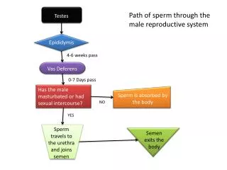
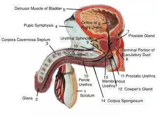
-pressure generated by the Sertoli cells pushes the sperm into a series of ducts within the testes that end up as the epididymis -within the epididymis is the ductus epididymis -also made up of a head, body and tail portion -site of sperm maturation – acquire mobility (14 days) -helps propel sperm into the: -vas (ductus) deferens: conducting tube from testis to urethra -connects to the tail of the epididymis -connects the testes to the urethra -made up of a pseudostratified columnar epithelium with a lamina propria connective tissue plus three layers of smooth muscle -contractions of these muscular layers propel the sperm Reproductive Ducts -spermatic cord supports the vas deferens + blood vessels (testicular artery and the pampiniform venous plexus), lymphatic vessels, the cremaster muscle and autonomic nerves -passes through the inguinal canal
ejaculatory duct – forms from the union of the seminal vesicle and the end of the vas deferens • pass through the prostate gland and terminate in the urethra • urethra: 3 sections: • A. prostatic - runs through the prostate • connects to ducts from the prostate and to the ejaculatory duct • B. membranous - between prostate and penis • -through the muscles of the perineum – urogenital diaphragm • C. spongy - through the erectile tissue of the penis
Male reproductive glands • -glands: seminal vesicles, prostate, bulbourethral glands • -produce fluid that combine with sperm to make semen • -semen: alkaline, activates sperm cells • 1. prostate: surrounds the urethra -forms as an outgrowth of the urethra along with the bulbourethral glands • -secretes a thin, milky fluid that enhances sperm motility and neutralizes vaginal fluid • 2. seminal vesicles: connect to urethra via the ejaculatory ducts • -secretes an alkaline fluid that contains sugars and prostaglandins (stimulates uterine contractions) • 3. bulbourethral glands: 2 glands behind • the prostate • -secrete a fluid that lubricates the penis
-conveys urine and semen -body is found externally -body is comprised of two tissue types of erectile tissue surrounded by connective tissue A. corpus cavernosum - large spaces B. corpus spongiosum - smaller spaces -surrounds the urethra -root of the penis is attached to the pelvis -corpus spongiosum enlargens at the tip - glans penis (sensory receptors) -glans penis covered with a loose fold of skin = prepuce
-ovary: production of egg -surface is covered with a germinal epithelium (simple epithelium) – does NOT give rise to the ova! -next layer is = tunica albuginea – dense irregular connective tissue capsule -outer cortex- granular tissue due to the presence of tiny ovarian follicles - inner medulla - connective tissue with blood & lymphatic vessels and nerves
Oogenesis and Follicular Development • begins before birth • early fetal development – primordial germ cells from the yolk sac migrate into the developing ovaries • differentiate to form oogonia (diploid stem cells) • undergo mitosis to produce millions of germ cells • most of the germ cells degenerate by atresia • a few develop further into primary oocytes – entered prophase I of meiosis • surrounded by a layer of follicular cells = primordial follicle • at birth – 200,000 to 2,000,000 primary follicles within the ovary • at puberty 40,000 are still present • 400 will develop further (rest undergo atresia) • -release of FSH and LH each month causes the development of one primary oocyte into a secondary oocyte • -development of a few primordial follicles into primary follicles (only one will continue until ovulation) • -primary follicle – primary oocyte surrounded by several layers of epithelial cells called granulosa cells • -develops a clear glycoprotein layer between the oocyte and the granulosa cells – zona pellucida • -the outermost granulosa cells contact a basement membrane which begins to develop into two layers (theca layers) • -now known as the secondary follicle • -secondary follicle begins to accumulate fluid in the center of the follicle (antrum) • -innermost granulosa cells firmly attaches to the zona pellucida = corona radiata • -becomes larger and turns into the tertiary or mature Graafian follicle • -completes meiosis I – two haploid cells • -these haploid cells are uneven in size but each have 23 chromosomes (two chromatids each - 46) • -smaller cell – first polar body (discarded nuclear material) • -larger cell – secondary oocyte • -receives most of the cytoplasm and has 23 chromosomes • -stops at metaphase II • -ovulated
Oogenesis and Follicular Development • ovulation – expulsion of the secondary oocyte into the pelvic cavity with the first polar body and corona radiata • fertilization – union of egg and sperm • penetration of the sperm into the secondary oocyte results in the resumption of meiosis II • the secondary oocyte splits again into two cells of unequal size (n) • larger one is called the ovum and the smaller one is the second polar body • combination of the ovum and the sperm results in the formation of the zygote • the first polar body splits also into two haploid cells • therefore meiosis of the primary oocyte produces one haploid ovum and three haploid polar bodies that degenerate
uterus: receives and nourishes the embryo • -comprised of a body, a curved portion (fundus) and the cervix • -uterine wall outer perimetrium, muscular myometrium and inner endometrium • -endometrium: mucosal layer covered with epithelium • -rich blood supply, sloughed off during menstruation • uterine tubes (Fallopian tubes): conduction of egg from ovary to uterus • -expands at end near the ovary = infundibulum with fimbrae (fingers) for the “catching” of the released egg • -lined with a mucosal layer and columnar epithelium with cilia -are also cells with microvilli rather than cilia – produce a nutritive fluid for the egg • cervix: projects into the vaginal canal
Female Reproductive Cycle • two cycles • 1. ovarian: during and after the maturation of the oocyte • 2. uterine: concurrent series of changes in the endometrium of the uterus to prepare it for embryo implantation
-3 major types of estrogens: • beta-estradiol • estrone • estriol • other smaller quantities • -follicular estrogens: • promote the development of the • female reproductive structures, • secondary sex characteristics and • the mammary glands • b. increase protein anabolism, including • bone synthesis • c. lower blood cholesterol • d. inhibit the release of GnRH, FSH • and LH -GnRH causes release of FSH and LH from anterior pituitary -FSH initiates follicular growth -LH stimulates the maturation of follicles -both LH and FSH stimulate the secretion of estrogens from the follicle -LH stimulates the theca layers of the follicle to make androgens -FSH stimulates the uptake of these androgens and converts them to estrogens -LH triggers ovulation and results in development of corpus luteum -corpus luteum produces and releases progesterone and some estrogen plus relaxin and inhibin -estrogen and progesterone regulate pregnancy, menstruation, secondary sex char’s & development of sex organs at puberty -relaxin – relaxes the uterus by inhibiting contractions of the myometrium -important to the implantation of the embryo – produced by the placenta during pregnancy -also increases the flexibility of the pubic symphysis -inhibin - inhibits secretion of FSH and LH
Menstrual Phase • Ovarian events – FSH increase causes • development of primordial follicles into • primary follicles • -may take several months to complete! • B. Uterine events – 50-150 mL of blood, • tissue fluid, mucus and epithelial cells • -shed from the stratum functionalis • -occurs because of declining levels of • E and P = causes the spiral arterioles to • contract which kills the cells of the SF • leaving the stratum basalis intact • Preovulatory Phase – most variable in length • A. Ovarian events – secretion of E and inhibin from the secondary follicles • -by day 6 one secondary follicle has outgrown the rest to become the dominant follicle • -the dominant follicle secretes E and I which causes an inhibition of FSH and a decrease in the stimulation of other follicles • -the dominant follicle develops into the Graafian follicle • -forms a blister-like bulge due to an increase in fluid within the antrum of the follicle • -the GF continues to increase its estrogen production • B. Uterine events – E stimulates the repair of the SF by stimulating mitosis in the SB layer • -the arterioles begin to lengthen and coil within the SF (4-10mm in thickness)
3. Ovulation • A. Ovarian events – rupture of the GF usually around day 14 • ovulated secondary follicle remains surrounded by its corona radiata and its zone pellucida • triggered by a positive feedback system – high levels of E at the end of the preovulatory phase increases the secretion of GnRH, also increases the release of LH directly • increased LH induces rupture of the GF about 9 hours after the LH peak • basis for the at-home ovulatory tests – detect rises in LH • B. Uterine events - none • signs of ovulation • Increase in basal body temperature • Changes in cervical mucus • Cervix softens • Mittelschmerz---pain
4. Postovulatory Phase – most consistent part of the cycle (14 days) • A. Ovarian events – the mature GF collapses and bleeding from the follicle results - the development of a blood clot results as the follicle induces bleeding – follicle is now called the corpus hemorrhagicum • the basement membrane between the granulosa cells and the thecal layers degenerates • this mixes the granulosa and thecal cells – transformed into corpus luteum cells under the influence of LH • luteal cells produce hormones and absorb the blood clot • if the ovum is NOT fertilized, the CL degenerates into the corpus albicans – 2 weeks • decrease in P, E and I results in the release of GnRH, FSH and LH (loss of negative feedback) • new follicular growth begins • if fertilized – the CL persists beyond 2 weeks by the secretion of human chorionic gonadotropin (hCG) hormone produced by the developing chorion that surrounds the embryo (8 days post-fertilization) • B. Uterine events – P and E produced by the corpus luteum promotes the growth and vascularization of the endometrium and its thickening to about 12-18 mm • -endometrial glands within the endometrium begin to secrete glycogen – energy for the fertilized egg
Surgical Hormonal Mechanical barriers Periodic abstinence Coitus interruptus Induced abortion Birth Control Methods
Reproductive disorders • Males • testicular cancer • prostate concer • erectile dysfunction (ED) • benign prostatic hyperplasia • Females • PMS • PMDD • Endometriosis • Ovarian, uterine cysts • Ovarian, uterine, cervical cancer • vulvovaginal candidiasis • Both • UTI • STDs – gonorrhea, syphillis, chlamydia, genital herpes, genital warts
Pregnancy -fertilization in the upper third of the oviduct/fallopian tube -fertilization = union of egg and sperm -plasma membrane of the egg is surrounded by an extracellular matrix = zona pellucida and a ring of follicular cells = corona radiata (nourishment in the follicle) -after fertilization = zygote 1. sperm penetrates corona radiata 2. several sperm enter zona pellucida -one of the glycoproteins within the ZP (ZP3) acts as a receptor for the sperm -binding causes dissolution of the acrosome and release of digestive enzymes 3. ONE sperm penetrates the plasma membrane of the egg 4. immediate change in the oocyte cell membrane (depolarizes) -also binding results in release of intracellular calcium which stimulates exocytosis of secretory vesicles whose contents inactivate ZP3 and harden the zona pellucida - impervious to more sperm 5. oocyte releases the zona pellucida away from the egg surface 6. fusion of the sperm with nucleus of the egg -before fusion the secondary oocyte must complete meiosis II and form the ovum
embryonic stage: week 2 to week 8 -after sperm penetration and ovum development the nuclei of the egg and sperm undergo changes to become pronuclei -union of sperm and egg pronuclei nuclei forms the zygote -first cell division = embryo -first division takes place 24 hours post-fertilization – takes 6 hours to complete -each succeeding division takes less time -72 hr stage = 16 cells -96 hr stage = morula (embryo is the size of the original ovum, filled with cells (blastomeres)
Implantation • attaches after about 6 days • usually in the fundus or the body of the uterus • orients its inner cell mass toward the uterus • 7 day – the endometrium becomes more vascularized • 9 days – completely embedded • following implantation, the endometrium is called the decidua • several layers with defined functions
-day 4 – formation of morula and passage into the uterine cavity -endometrial glands release a glycogen-rich fluid = uterine milk -enters the morula through the zona pellucida and provides nourishment -day 5 -the fluid begins to collect in the morula and reorganizes them around a fluid-filled cavity = blastocoel -embryo is now called a blastula or blastocyst (50-150 cells) -outer layer = trophoblast - forms extraembryonic tissues (e.g. placenta, yolk sac) -inner cell mass at one end - totipotent embryonic stem cells -by the end of day 5, the blastocyst digests a hole in the ZP and squeezes through it to undergo implantation
-second week of development - the inner cell mass flattens = embryonic disk (hypoblast and epiblast) -hypoblast = primitive endoderm -epiblast = primitive ectoderm -amniotic cavity forms between the inner cell mass and the trophoblast -surrounded by an amniotic membrane – develops from the epiblast -fills with amniotic fluid – filtrate from maternal blood at initial stages -formation of the yolk sac (from the hypoblast) -forms blood cells, gives rise to sex cells and the stem cells of the immune system -also forms part of the embryonic digestive tube -portion will also become part of the umbilical cord -the outer trophoblast cells develops into two layers within the region where the blastocyst and the endometrium make contact – become part of the chorion -these trophoblast cells will secrete digestive enzymes that allow the embryo to burrow into the decidua -also secrete hCG – rescues the corpus luteum from degeneration
day 15: embryonic disk undergoes gastrulation to form the gastrula embryonic stage • formation of the three embryonic germ layers by differentiation of the ES cells within the embryonic disc • epiblast form a specialized region = primitive streak • clearly establishes a head and tail orientation • head end the streak enlargens to form the primitive node • cells from the epiblast move inward through the primitive streak • some cells displace the hypoblast and form the endoderm • other cells are retained in the area and form the mesoderm • mesoderma forms a loose connective tissue = mesenchyme • cells remaining in the epiblast form the ectoderm
-portions of the mesoderm that do not form the notochord segment into sections called somites -> specific body regions and structures • -in front of the primitive streak forms the primitive node – head and associated structures • mesodermal cells from the primitive node form a hollow tube near the future head of the • embryo - become the notochord (day 22-24) • (progenitor to the vertebral column) • -four weeks of development - embryo forms a tubular structure • -embryo begins to form definitive structures: • -neural folds of ectoderm -> nervous system (brain and spinal cord) • ** neurulation occurs by induction (one tissue influences the development of another) • -e.g. nervous system requires the mesodermal cells of the notochord
Medical application: Stem Cells • two broad categories of mammalian stem cells exist: embryonic stem cells, and adult stem cells • Stem cells = primal cells that: • 1) retain the ability to renew themselves through cell division and 2) can differentiate into a wide range of specialized cell types • stem cell field grew out of findings by Canadian scientists Ernest A. McCulloch and James E. Till in the 1960s • 1963 – self-renewing cells in the bone marrow of mice • 1964 - single cells from a human testicular teratocarcinoma were isolated • - remained undifferentiated in culture = embryonic carcinoma cells (EC cells)
1968 – bone marrow transplant was used to cure two patients of SCID • 1978 – Hematopoietic Stem Cells (HSCs) were isolated from the bone marrow and identified • 1981 – Embryonic stem cells (ES cells) first isolated and cultured from mouse embryos in 1981 by two independent research groups. Evans and Kaufman, Martin • 1998 - human embryonic stem cells isolated and grown by James Thomson at the University of Wisconsin-Madison • developed a technique to isolate and grow ES cells from human blastocysts
stem cells can be defined by their ability to form specific cell types • potency = ability to specialize into a distinct cell type • -unipotent – ability to form only one cell type • e.g. pre-adipocyte -> adipocyte • sometimes called progenitor cells because of their limited ability to specialize • -bipotent – ability to form two cell types • e.g. osteochondro progenitor cells (OPCs) – bone and cartilage • -multipotent/pluripotent – ability to form many cell types • multipotent – usually within one germ lineage (mesoderm) • e.g. MSC & ASCs – bone, fat, cartilage and muscle • pluripotent – within more than one lineage (mesoderm & ectoderm) • -adult stem cells???? • -totipotent – ability to form all cell types • e.g. ES cell
adult stem cell populations • bone marrow – HSC and MSC • adipose – ASC • skin • brain/neural - NSCs • skeletal muscle • mammary • olfactory • hepatic • corneal • etc……. • fetal stem cell populations – placental and fetal tissue derived • -placental & amniotic populations • -umbilical cord blood • increased potentials because of their “younger” age??? • adult stem cells may have limited potency when compared to ES cells • but are still useful because of their limited ethical concerns • in addition ES cells still may possess the ability to form cancers • -many proto-oncogenes are involved in embryonic and fetal development • -these genes turn on and off at specific times during development • -if they turn back on during adult stages = oncogenes – may cause the formation of tumors • http://stemcells.nih.gov/
Use of adult stem cells • use of bone marrow stem cells (i.e. HSCs) leukemia and lymphoma • Potential treatments • 2.1 Brain Damage • neural stem cells • rats subjected to stroke - administration of drugs to increase the NSC division rate and may increase the survival and differentiation of newly formed cells • within weeks, recovery of brain structure is accompanied by recovery of lost limb function • several studies in which NSCs or ES cells are injected into damaged areas of the brain • treatment for Parkinson’s • 2.2 Cancer • 2.3 Spinal cord injury • University of Wisconsin-Madison: differentiated human blastocyst stem cells into neural stem cells, then into the beginnings of motor neurons, and finally into spinal motor neuron cells • University of California: injection of hES cells into paralyzed mice – limited regaining of their ability to move and walk four months later. • stem cells regenerated neurons in addition to the myelin sheath • http://img227.imageshack.us/img227/7954/stemcellbreakthru052wl.jpg • 2.4 Muscle damage - use in muscular dystrophy and myasthenia gravis • 2.5 Heart damage – repair of ischemic coronary arteries • 2.6 Low blood supply • 2.7 Baldness • stem cells within hair follicles • follicle stem cells may lead to successes in treating baldness through "hair multiplication," also known as "hair cloning," as early as 2008???? • 2.8 Missing teeth • 2.9 Deafness • 2.10 Blindness and Vision Impairment • 2.11 ALS (Lou Gehrig's Disease) • Johns Hopkins University: induced nerve damage similar to that of ALS • injection of rats with stem cells - migration to the sites of injury • regeneration of the dead nerve cells • restoration of movement • (http://www.cellmedicine.com/als.asp)
Therapeutic cloning • In somatic cell nuclear transfer (SCNT) the nucleus of a somatic cell is removed and the rest of the cell is discarded. • the nucleus of an egg cell is removed • the nucleus of the somatic cell is then inserted into the enucleated egg cell. • the egg is stimulated to divide by an electric shock (depolarizes the egg’s plasma membrane) • new cell begins to divide and proceedes through the various embryonic stages • SCNT is used in stem cell research - to obtain stem cells that are genetically matched to the donor organism • e.g. potential use of genetically-customized stem cells & Parkinson's disease - stem cells resulting from SCNT would those genes that contribute to Parkinson's disease. therefore, the disease-specific stem cell lines could be studied in order to better understand the disease • e.g. genetically-customized stem cell lines could be generated for cell-based therapies to transplant to the patient - avoiding any complications from immune system rejection • *** no human stem cell lines have been derived from SCNT research. In 2005, a South Korean research team led by Professor Hwang Woo-suk, published claims to have derived stem cell lines via SCNT ,but supported those claims with fabricated data
Cloning types • Molecular cloning • procedure of isolating a DNA sequence of interest and obtaining multiple copies of it in an organism. • frequently employed to amplify DNA fragments containing genes, an essential step in their subsequent analysis. • cloning of any DNA sequence involves the following four steps: • fragmentation, ligation, transfection, and screening/selection. • Genetic cloning • Cloning a cell means to derive a (clonal) population of cells from a single cell. Asexual reproduction (also known as agamogenesis) is a form of reproduction which does not involve meiosis, gamete formation, or fertilization. • In laymen's terms, there is only one "parent" involved. • common among simple organisms such as amoeba and other single-celled organisms, • Horticultural • -clone in horticulture means all descendants of a single plant, produced by vegetative reproduction • -many horticultural plant cultivars are clones - multiplied by some process other than sexual reproduction. • e.g. some European grapes represent clones that have been propagated for over two millennia. • - other examples are potato and banana. • -Grafting can be regarded as cloning, since all the shoots and branches coming from the graft are genetically a clone of a single individual • -Many trees, shrubs, vines, ferns and other herbaceous perennials form clonal colonies.
Reproductive Cloning • SCNT can also be used in the reproductive cloning of animals (e.g. Dolly the sheep), and in theory could be used to clone humans. • embyro is created by SCNT & transferred to the uterus of a female host where it continues to develop until birth • Dolly or any other animal created using nuclear transfer technology is not truly an identical clone of the donor animal. • Only the clone's chromosomal or nuclear DNA is the same as the donor. • the mitochondrial DNA will differ • mutations will occur throughout embryonic and fetal development • so the clone is not 100% identical! • Species cloned • Tadpole: (1952) Many scientists questioned whether cloning had actually occurred and unpublished experiments by other labs were not able to reproduce the reported results. • Carp: (1963) In China, embryologist Tong Dizhou cloned a fish. He published the findings in an obscure Chinese science journal which was never translated into English.[1] • Mice: (1986) first successfully cloned mammal; Soviet scientists Chaylakhyan, Veprencev, Sviridova, Nikitin had mice "Masha" cloned. • Sheep: (1996) from early embryonic cells by Steen Willadsen. Megan and Morag cloned from differentiated embryonic cells in June 1995 and Dolly the sheep in 1997. • Rhesus Monkey: Tetra (female, January 2000) from embryo splitting • Cattle: Alpha and Beta (males, 2001) and (2005) Brazil • Cat: CopyCat "CC" (female, late 2001), Little Nicky, 2004, first cat cloned for commercial reasons • Mule (2004): Idaho Gem, a john mule - the first horse-family clone. • (2003) Horse: Prometea • a similar process called budding has been used in cattle for decades • -an embryo is dissociated into individual cells without harm • -each cell – separate embryo • -animal is not derived from a differentiated cell but from a undifferentiated egg
HIV and AIDS -most viral illnesses are caused by DNA viruses a. binding of virus to host cells via interaction of host and viral envelope proteins - endocytosis b. degradation of viral capsid proteins to release the naked viral DNA c. viral DNA enters host nucleus and “takes over” host machinery - d. replication, transcription and translation of viral components e. assembly of new viral capsids and formation of new viral progeny f. release of progeny from host cell (cell death of host)
HIV • Previous names: • human T-lymphotropic virus-III (HTLV-III) • lymphadenopathy-associated virus (LAV) • AIDS-associated retrovirus (ARV) • Infection - transfer of blood, semen, vaginal fluid, pre-ejaculate or breast milk. • HIV is present as both free virus particles and virus within infected immune cells. three major routes of transmission: • unprotected sexual intercourse • contaminated needles and • transmission from an infected mother to her baby HIV primarily infects vital cells of the immune system • HIV primarily infects vital cells of the immune system • e.g. helper T cells (specifically CD4+ T cells), macrophages and dendritic cells • infection leads to low levels of CD4+ T cells through three main mechanisms: • 1. direct viral killing of infected cells • 2. increased rates of apoptosis in infected cells • 3. killing of infected CD4+ T cells by CD8 cytotoxic lymphocytes – recognize infected cells. • when CD4+ T cell numbers decline below a critical level – loss of cell-mediated immunity (T cell mediated immunity) • body becomes progressively more susceptible to opportunistic infections. • about one in ten remain healthy for many years, with no noticeable symptoms
HIV timeline • AIDS epidemic was discovered June 5, 1981 • U.S. Centers for Disease Control and Prevention reported a cluster of Pneumocystis carinii pneumonia in five homosexual men in Los Angeles • originally dubbed GRID, or Gay-Related Immune Deficiency • 1982 - the CDC introduced the term AIDS • 1983 - scientists led by Luc Montagnier at the Pasteur Institute in France discovered the HIV • originally called lymphadenopathy-associated virus (LAV). • 1984 - a team led by Robert Gallo of the US confirmed the discovery of the virus • renamed it human T lymphotropic virus type III (HTLV-III) • President Mitterrand and President Reagan had to resolve the discovery issues • 1986 - renamed human immunodeficiency virus (HIV) • Three of the earliest known instances of HIV-1 infection are as follows: • 1. A plasma sample taken in 1959 from an adult male living in what is now the Democratic Republic of Congo • 2. HIV found in tissue samples from a 15 year old African-American teenager who died in St. Louis in 1969 • 3. IV found in tissue samples from a Norwegian sailor who died around 1976
HIV classification • HIV classified as a member of the genus lentivirus - part of the family of retroviridaeLentiviruses: responsible for long-duration illnesses, long incubation period • transmitted as single-stranded, positive-sense, enveloped RNA virus • upon entry of the target cell, the viral RNA genome is converted to double-stranded DNA by a virally encoded reverse transcriptase • viral DNA is then integrated into the cellular DNA by a virally encoded integrase after cell infection - two pathways are possible: • 1. either the virus becomes latent and the infected cell continues to function • 2. the virus becomes active and replicates – produces a large number of progeny viruse particles • Two species of HIV infect humans: HIV-1 and HIV-2. • HIV-1 is thought to have originated in southern Cameroon after jumping from wild chimpanzees (Pan troglodytes troglodytes) to humans • HIV-1 is more virulent. • easily transmitted • is the cause of the majority of HIV infections globally. • HIV-1 is the virus that was initially discovered and termed LAV. • HIV-2 may have originated from the Sooty Mangabey (Cercocebus atys), an Old World monkey of Guinea-Bissau, Gabon, and Cameroon • HIV-2 is less transmittable • largely confined to West Africa
Structure and genome • HIV is different in structure from other retroviruses. • approximately 120 nm in diameter (around 60 times smaller than a red blood cell) roughly spherical. • composed of two copies of positive single-stranded RNA • RNA codes for the virus's nine genes – but it must be converted back into DNA (reverse transcribed by an enzyme called reverse transcriptase) • enclosed by a conical capsid composed of 2,000 copies of the viral proteinp24 • single-stranded RNA is tightly bound to: nucleocapsid proteins, p7 and enzymes needed for the development of the virion such as reverse transcriptase, proteases, ribonuclease and integrase • the capsid is surrounded by a matrix composed of the viral protein p17 • ensures the integrity of the virion particle • matrix is surrounded by a viral envelope - two layers phospholipids taken from the membrane of a human cell upon budding of the progeny virsus from its host • viral envelope is embedded with proteins from the host cell and about 70 copies of a complex HIV protein called Env • Env - cap made of three molecules called glycoprotein (gp) 120, and a stem consisting of three gp41 molecules (anchor the Env complex to the viral envelope) • enables the virus to attach to and fuse with target cells • both gp120 and gp41 (especially gp120) are targets of future treatments or vaccines • 3 genes = gag, pol, and env contain information needed to make the structural proteins for new virus particles • 1. env - codes for gp160 which is processed to form gp120 and gp41 • that is broken down by a viral enzyme to form gp120 and gp41 • 2. gag– codes for the capsid protein 024, the nucleocapsid proteins p6, p7 and the matrix protein p17 • 3. pol – codes for the enzymes reverse transcriptase, integrase and protease • the six remaining genes are tat, rev, nef, vif, vpr, and vpu (or vpx in the case of HIV-2) - are regulatory genes that code for proteins that control the ability of HIV to infect cells, replicate, or cause disease • e.g. nef appears necessary for the virus to replicate efficiently, and the • e.g. vpu influences the release of new virus particles
HIV Tropism • viral tropism = cell types HIV infects. • HIV infects a variety of immune cells such as CD4+ T cells, macrophages, and microglial cells • HIV-1 entry to macrophages and CD4+ T cells is mediated through interaction of gp120 with: • 1. the CD4 molecule • 2. with chemokine coreceptors • Strains of HIV-1 • 1. Macrophage (M-tropic) or non-syncitia-inducing strains (NSI) • use the β-chemokine receptor CCR5 for entry • able to replicate both in macrophages and CD4+ T cells • CCR5 receptor is used by almost all primary HIV-1 isolates regardless of viral genetic subtype. • macrophages appear to be the first cells infected by HIV • perhaps the source of HIV production when CD4+ cells become depleted in the patient • microglial cells in the CNS can be infected by the NSI strains • 2. T-tropic isolates, or syncitia-inducing (SI) strains • replicate in primary CD4+ T cells and macrophages • use theα-chemokine receptor, CXCR4 • administration of SDF-1(a ligand for CXCR4) may be able to suppress replication of T-tropic HIV-1 isolates by down-regulating the expression of CXCR4 on the surface of these cells • HIV strains that use only the CCR5 receptor are termed R5 • those that only use CXCR4 are termed X4 • those that use both = X4R5 • Some people are resistant to certain strains of HIV • e.g. people with a mutation in the CCR5 gene (CCR5-Δ32 mutation); these resistant to infection with R5 virus - HIV cannot bind this coreceptor thus reducing its ability to infect target cells. • Both X4 and R5 HIV are present in the seminal fluid which is passed from partner to partner • R5 strain seems to predominate • unknown why – but spermatozoa may selectively carry R5 HIV • they possess both CCR3 and CCR5 but not CXCR4 on their surface • genital epithelial cells preferentially infected by the X4 virus