Download

1 / 28
290 likes | 439 Views
Dynamics of Blood Flow. 5.7.12. R 1. R 2. R 3. R 1 ,Q 1. D P 1. D P 2. D P 3. R 2 ,Q 2. D P= D P 1 + D P 2 + D P 3 =QR 1 +QR 2 +QR 3 =QR R=R 1 +R 2 +R 3. Q=Q 1 +Q 2 = D P/R 1 + D P/R 2 = D P/R 1/ R=1/R 1 +1/R 2.

E N D
Dynamics of Blood Flow 5.7.12
R1 R2 R3 R1,Q1 DP1 DP2 DP3 R2,Q2 DP= DP1 + DP2 + DP3 =QR1+QR2+QR3 =QR \R=R1+R2+R3 Q=Q1+Q2 =DP/R1+DP/R2 =DP/R \1/R=1/R1+1/R2
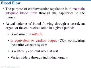
The slow velocity of blood through capillaries has the beneficial effect of allowing more time for the exchange of molecules between the cardiovascular system and extracellular fluid
Blood flow through stenosis • Bernoulli’s principle states that when the fluid flow through a tube is constant, the total fluid energy –the sum of kinetic energy and potential energy-remains constant • It explains why fluid pressure is low in blood vessels at places where its radius is less
Blood flow through stenosis • Proximal to the structure of stenosis, blood flow is laminar. Blood passing through stenotic orifice also remains laminar, but its velocity increases to maintain the volumetric rate of flow. Here the pressure against the walls is least and hence there is a pressure drop across the narrower area of a vessel
Volume of blood contained within a vessel • The volume of blood contained within a flexible blood vessel depends on • Transmural Pressure: • Degree of flexibility within the vessel wall • Transmural pressure is always defined as the difference in pressure inside versus outside a hollow structure
Vasscular distensibility is expressed as the fractional increase in volume for each mmHg rise of pressure • All the vessels though the extent varies are distensible (flexible) • Distensibility depends on Vessel wall tension Elastic properties of vessel constituent materials
Distensibility and Elasticity • Vessels which are highly elastic are called Windkessel vessels such as aorta and its large branches. • Conducting arteries (conducting because they offer much less resistance to blood flow due to large diameter) i. e. aorta and its major branches are most elastic vessels because they contain more elastin than any other vessel type
Distensibility and Elasticity • These vessels store part of the energy produced by cardiac ejection as potential energy and in doing so their walls are distended during systole. During diastole recoil of the walls converts stored energy into kinetic energy for circulation • Elastic recoil of the arterial system helped by the resistance to outflow offered by peripheral arterioles convert the pulsatile ejection of heart into a steady flow
Elastic recoil of arteries • The heart generates high pressure within the ventricles when it contracts during systole which then drops to near zero during a diastole • During diastole, elastic recoil of arteries pushes blood forward against the downstream vascular resistance generating a significant diastolic pressure • For this reason diastolic pressure drops to only about 80mmHg in the aorta as compared with near zero in ventricles
Veins are most distensible vessels. Veins by changing their luminal configuration i.e. from elliptical to circular cross sectional profiles adjust the capacity of vascular system
Vascular Wall Tension • Any transmural pressure within a vessel exerts a force on the vessel wall that would tend to rip the wall apart were it not for the opposing force supplied by the muscle and connective tissue of the vessel wall • This opposing force is called the wall tension • The tension T is roughly proportional to transmural pressure i.e. pressure difference on the inside and outside of the wall and the radius r of the vessel. Thus T = a P r Here ‘a’ is a constant. This is Laplace law
Vascular Wall Tension • More precisely T= P r /w where w is the thickness of the vessel wall • Small vessels are able to withstand higher pressures than vessels of larger diameter
Vascular Wall Tension • Capillaries with small diameter can withstand relatively high intravascular pressures even though they are composed of a single layer of endothelial cells • In arteries and veins, vessels with thick walls relative to their radius are able to withstand high pressure than vessels with small r/w rato
Volume of Blood within vessels • Large transmural pressure within flexible vessels create large intravascular volumes • Small transmural pressure in stiff walled vessels produce small intravascular volumes Although arteries are quite distensible because they are most elastic but elastic recoil of arterial wall decreases he amount of blood contained within them
Vascular Compliance • Vascular compliance is given by • The compliance of vein is about 24 times that of its corresponding artery
Special features of Blood Flow Fahreus-Lindqvist Effect: • Relative viscosity of water, serum or plasma is not altered when they are made to flow through tubes of different sizes • But the relative viscosity of blood is altered when it passes through tubes of different sizes i.e. blood flow in very minute vessels exhibit far less viscous effect than it does in large vessels. This is called Fahreus-Lindqvist Effect • in particular there's a decrease of viscosity of blood as it moves from larger vessels to smaller ones (only if the vessel diameter is between 10 and 300 micrometers). • The viscosity decreases with decreasing capillary radius r. This decrease was most pronounced for capillary diameters < 0.5mm
Fahreus-Lindqvist Effect • Reasons for Fahreus-Lindqvist Effect: Segre–Silberberg effect, Plasma Skimming Change in haematocrit
Plasma Skimming • For deformable particles (such as red blood cells) flowing in a tube, there is a net hydrodynamic force that tends to force the particles towards the center of the vessels with diameter between 10 and 300 micrometers This is known as the Segre–Silberberg effect. • On average, there will be more red blood cells near the center of the capillary than very near the wall, leaving plasma at the wall of the vessel • There is a cell free layer near the vessel wall. This redistribution is known as “plasma skimming”
In larger vessels such as aorta and its major branches, the cell free layer is only a small % of complete blood stream, so it does not effect the viscosity of blood much • However in arterioles (<300µm) and capillaries, this layer becomes a great % of the total volume contained within the vessel, so fluid velocity as a whole decreases in these vessels
Plasma Skimming • Newtons law of motion under translational and rotational motion • If the red blood cell is not located directly within the flow centerline (assuming that the flow is fully developed and parabolic, then the forces induced by the fluid velocity will be different on each end of the red blood cell. Due to this influence of forces, the red blood cell will tend to rotate towards the higher flow region • Recall that the higher fluid velocity has a lower shear stress and therefore higher shear stress would be localized closer to the vessel wall
Plasma Skimming • This continues until the forces acting o the red cell are balanced and do not cause rotation • Therefore red cells move to the lower shear stress region which have high velocities in order to balance forces.
Change in haematocrit • Another reason for Fahreus-Lindqvist Effect is change in haematocrit as the vessel diameter decreases • The haematocrit in capillaries e.g. is lower than in the arteries. This is because red blood cells are fairly restricted to the centerline (higher velocity flow) in capillaries and plasma is slowest at the vessel wall • With an inflow haematocrit of 40-50%, capillary haematocrit is about 10-20%
Plug flow • Red blood cells in the capillaries tend to plug the capillary preventing the plasma to flow freely as it does in large blood vessels • While the red blood cells flow fairly steadily, the plasma in between the cells experiences turbulent eddies and recirculation zones • These eddies helps to move materials from the centerline of the plasma towards the vessel wall, potentially helping in the transfer of materials across the capillary wall
Plug flow • These turbulent eddies tend to move compounds to the endothelial cell wall, so that they can diffuse across the wall, instead of convect across the blood and then diffuse across the wall • There is a very small layer of plasma along the blood vessel wall that experiences very high shear stresses, because it is being squeezed between a red blood cell and an endothelial cell. Here the shear stress increases significantly
55 mm Hg -35 mm Hg 100 mm Hg 1 mm Hg 95 mm Hg 100 mmHg 95 mmHg 195 mm Hg 105 mm Hg Effect of gravity on Blood Pressure Venous pressures • Effect of gravity on pressure • Distance heart-head~ 0.4 m • Heart-feet ~ 1.4 m • DP = rgh Arterial pressures
Effect of gravity on Blood Pressure • The pressure in any vessel above heart level is decreased by the effect of gravity • The arterial pressure is increased by 0.77mmHg for every centimeter below the right atrium and similarly decreased for each cm above the right atrium