Download

1 / 45
450 likes | 565 Views
The Kidney Part Three – The Renal Corpuscle Digital Laboratory. It’s best to view this in Slide Show mode, especially for the quizzes. This module will take approximately 45 minutes to complete .

E N D
The Kidney Part Three – The Renal Corpuscle Digital Laboratory It’s best to view this in Slide Show mode, especially for the quizzes. This module will take approximately 45 minutes to complete.
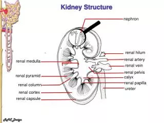
After completing this exercise, you should be able to (blue text in this module, tan text in subsequent kidney modules): • describe the gross anatomical features of the kidneys • Cortex • Medulla, including renal pyramid, renal papilla, renal columns • Hilus, sinus, renal pelvis, and major and minor calyces • recognize and discriminate between the pars convoluta and the pars radiata. • diagram blood circulation through the kidneys, and identify the major renal vessels on a histological section: • Renal artery and vein • Interlobar artery and vein • Arcuate artery and vein • Interlobular artery and vein • Afferent arteriole, glomerulus, efferent arteriole • Peritubular capillaries and vasa recta. • distinguish, at both the light and electron microscopic level, each of the following renal tubular structures: • proximal convoluted tubule • thick descending limb of the loop of Henle • thin limb of the loop of Henle • thick ascending limb of the loop of Henle • distal convoluted tubules • collecting (connecting) ducts, papillary ducts • Identify glomeruli at the light microscopic level, as well as identify each of the following in an electron micrograph: • endothelial cells • podocytes, including their primary and secondary pedicels (foot processes) • filtration slits • parietal epithelium • lamina densa • mesangial cells • blood space • urinary space. • Identify a juxtaglomerular apparatus, including the macula densa and juxtaglomerular cells
formation of the renal corpuscle To understand the final structure of the renal corpuscle, it is useful to think about the end of the nephron during development. Blood vessels in the connective tissue form a tuft of capillaries, the glomerulus, flanked by two arterioles (afferent and efferent). The afferent arteriole is a branch of an interlobular artery, while the efferent arteriole feeds the peritubular capillary plexus. Note the endothelial cells of the glomerulus have a basement membrane (not shown). Connective tissue Basement membrane Epithelial cells Lumen of tubule (continuous with outside world)
formation of the renal corpuscle The renal tubule invaginates, forming a visceral and parietal layer of Bowman’s capsule. Cells of the visceral layer, called podocytes, cover the endothelial cells of the glomerulus. In many places, the connective tissue between the endothelial cells and podocyes is squeezed out, and the two basement membranes fuse to form a single, thick basal lamina, called the lamina densa. Green represents connective tissue
formation of the renal corpuscle Really cool 3D views of the corpuscle, with blood vessels in red, the visceral layer looks like saran wrap. Areas where basal lamina of endothelial cells and podocytes are fused. The connective tissue in these locations is called mesangium, the cells called mesangial cells.
the renal corpuscle - Light micrographs Light micrograph of a corpuscle. The tuft in the center contains the glomerulus, podocytes, and mesangial cells. The outer layer is the parietal cells. Parietal cells podocytes/endothelial cells/mesangial cells (cannot distinguish between these three in light micrographs) Capsular space Blood flows through the glomerular capillaries at relatively high pressure (for capillaries), forcing fluid into the capsular space. This fluid is provisional urine (aka ultrafiltrate) that flows into the proximal convoluted tubule.
the renal corpuscle - Light micrographs • Some fortuitous sections of renal corpuscles demonstrate: • vascular pole –vessels enter and exit the corpuscle • urinary pole - proximal convoluted tubule drains capsular space Many sections of renal corpuscles show neither of these (see previous slide).
the renal corpuscle - Light micrographs Video showing corpuscles - SL116 • Link to SL 116 • Be able to identify: • Parietal cells • Capsular space • Vascular pole • Urinary pole
the renal corpuscle - ELECTRON micrographs • Although the structure of a corpuscle in light micrographs is fairly straightforward, important details are revealed in electron micrographs. This drawing reinforces key features of a corpuscle. • Endothelium of glomerulus • Podocyte of visceral layer of glomerular capsule • Parietal layer of glomerular capsule • Capsular space
the renal corpuscle - ELECTRON micrographs • Below is a detailed drawing and scanning electron micrograph of glomerular capillary, viewed from within capsular space. Note: • The capillary endothelium is fenestrated (without diaphragms) • Podocytes are elaborate cells, with numerous extensions called pedicels (aka foot processes) • The pedicels of adjacent podocytesinterdigitate; and the space between the pedicels are called filtration slits Serum components pass through the fenestrations, basement membrane, and filtration slits to reach the capsular space.
the renal corpuscle - ELECTRON micrographs • In this scanning electron micrograph, you are within the capsular (urinary) space, looking down on a podocyte. Note parts of podocyte: • “cell body” • 1o pedicels • 2o pedicels • Filtrationslits 1o pedicels are the initial processes of the podocyte, 2o pedicels are the terminal processes that line the filtration slits.
the renal corpuscle - ELECTRON micrographs Another scanning electron micrograph of a podocyte: The red dotted line indicates the direction of blood flow, the yellow line indicates provisional urine production and flow into the proximal convoluted tubule.
the renal corpuscle - ELECTRON micrographs The electron micrograph to the right is a section similar to the blue line in the above drawing. It is a complicated image, so we’ll do it piece-meal in the next few slides. The outlined region is the “tuft” of capillaries and surrounding podocytes in the center of the corpuscle. The outer layer of the corpuscle is the parietal layer, labeled here as BC for Bowman’s capsule. The capsular space is indicated as well, labeled BS for Bowman’s space.
the renal corpuscle - ELECTRON micrographs Within the tuft you can see the lumen of the capillaries, labeled C. The fenestrated endothelial cells line the capillary (E).
the renal corpuscle - ELECTRON micrographs All of the cell fragments surrounding the capillaries belong to podocytes, shown in fragments due to plane of sectioning: P indicates a podocyte nucleus P1 indicates a 1o pedicel P2 indicates 2o pedicels The basement membrane (BM) is situated between the endothelial cells and podocytes. Mesangial cells (M), and mesangial (extracellular) matrix (MM) are also indicated (more on those later).
the renal corpuscle - ELECTRON micrographs • Same image, with all labels indicated so you can put it all together: • BC = Bowman’s capsule (parietal layer) • BS = Bowman’s space (capuslar space) • P = podocyte • P1 and P2 = pedicils (1o and 2o) • C = capillary lumen • E = endothelial cell • F = fenestrations of endothelial cell • BM = basement membrane, actually basal lamina or lamina densa • M = mesangial cell; MM = mesangial matrix
the renal corpuscle - ELECTRON micrographs The drawing to the lower right will be used for orientation on the following slides. This drawing is similar to a cut through the glomerular capillaries as indicated by the blue line in the left image, but does not include the parietal layer. Therefore, the capsular space is all the space surrounding the capillaries/podocytesas indicated. Capsular space Capsular space Capsular space Capsular space Capsular space Capsular space
the renal corpuscle - ELECTRON micrographs • A magnified view of three loops of glomerular capillaries, note: • P = podocyte • P1 and P2 = pedicils (1o and 2o) • C = capillary lumen • E = endothelial cell • F = fenestrations of endothelial cell • BM = basement membrane The basement membrane is actually a basal lamina. Because it is very thick in the corpuscle, it is also referred to as the lamina densa. capsular space Blue box is meant for approximate perspective, and doesn’t reflect exact orientation of capillaries.
the renal corpuscle - ELECTRON micrographs • Further magnification focuses on the blood-urine filter. Note: • P2 = 2o pedicels • E = endothelial cell • F = fenestrations of endothelial cell • BM = basement membrane (lamina densa) Capsular space (provisional urine here) Capillary lumen (blood here)
the renal corpuscle - ELECTRON micrographs Some more challenging images of the blood-urine barrier: podocyte tangential cut of fenestrations Capsular space fp = foot processes of podocyte FS = filtration slit US = urinary space (capsular space) RBC Capsular space RBC
the renal corpuscle - ELECTRON micrographs Some more challenging images of the blood-urine barrier. RBC Capsular space RBC RBC Capsular space When trying to figure out which side is which, I try to look for blood vessels. If they aren’t there, I move on and look for podocytes with their pedicels.
the renal corpuscle - ELECTRON micrographs There is connective tissue between glomerular capillaries, called the mesangium. Cells within this connective tissue are mesangial cells. The fused basal lamina splits on either side of the mesangium, so that there is a basal lamina between the endothelial cells and the mesangium, and between the podocytes and mesangium (advance to see arrows) Green represents connective tissue
the renal corpuscle - ELECTRON micrographs • In this micrograph, note the fused basal lamina splits on either side of mesangium (purple and orange arrows indicate basal lamina that has split): • BM = basement membrane, actually basal lamina or lamina densa • MC = mesangial cell; MM = mesangial matrix • P = podocyte; PD = pedicels • E = erythrocyte • L = lymphocyte • EC = endothelial cell • US = urinary (capsular) space Blue box is meant for approximate perspective, and doesn’t reflect exact orientation of capillaries.
the renal corpuscle – Macula densa The distal convoluted tubule of the nephron loops toward the corpuscle and lies adjacent to the afferent arteriole at the vascular pole of the corpuscle. Cells of the distal convoluted tubule adjacent to the afferent arteriole are the macula densa. Cells of the afferent arteriole adjacent to the distal convoluted tubule are the juxtaglomerular cells. Together, these form the juxtaglomerular apparatus, which is involved in regulating blood volume by releasing renin.
the renal corpuscle – Macula densa Light micrograph showing macula densa cells (outlined in blue) of a distal convoluted tubule. The original image without the outline is to the right. What you are looking for is a distal convoluted tubule that is adjacent to the vascular pole of the corpuscle, and the macula densa cell nuclei “line up” adjacent to the corpuscle.
the renal corpuscle – Macula densa Light micrograph showing macula densa cells (yellow arrows) of a distal convoluted tubule. The smaller nuclei just to the left of the macula densa are likely to be either mesangial cells (lacis cells) or juxtaglomerular cells, but you won’t have to specifically identify these.
the renal corpuscle – Macula densa Video showing macula densa - SL116 • Link to SL 116 • Be able to identify: • macula densa
The next set of slides is a final quiz for this module. You should review the structures covered in this module, and try to visualize each of these in light and electron micrographs. • describe the gross anatomical features of the kidneys • Cortex • Medulla, including renal pyramid, renal papilla, renal columns • Hilus, sinus, renal pelvis, and major and minor calyces • recognize and discriminate between the pars convoluta and the pars radiata. • diagram blood circulation through the kidneys, and identify the major renal vessels on a histological section: • Renal artery and vein • Interlobar artery and vein • Arcuate artery and vein • Interlobular artery and vein • Afferent arteriole, glomerulus, efferent arteriole (on a typical section, you cannot distinguish between the afferent and efferent arteriole) • Peritubular capillaries and vasa recta. • distinguish, at both the light and electron microscopic level, each of the following renal tubular structures: • proximal convoluted tubule • thick descending limb of the loop of Henle • thin limb of the loop of Henle • thick ascending limb of the loop of Henle • distal convoluted tubules • collecting (connecting) ducts, papillary ducts • Identify glomeruli at the light microscopic level, as well as identify each of the following in an electron micrograph: • endothelial cells • podocytes, including their primary and secondary pedicels (foot processes) • filtration slits • parietal epithelium • lamina densa • mesangial cells • blood space • urinary space. • Identify a juxtaglomerular apparatus, including the macula densa and juxtaglomerular cells (juxtaglomerular cells are difficult to definitively identify)
quiz Self-check: Identify 1-6. (advance slide for answers)
quiz Self-check: Identify 1-6. (advance slide for answers)
quiz Self-check: Identify 1-7. Note 1 & 2 are part of low magnification view, rest of image is magnified view of what is within rectangle. (advance slide for answers)
quiz Self-check: Identify outlined structure. (advance slide for answers) Macula densa
quiz Self-check: Identify. (advance slide for answers) 1o pedicel of podocyte 2o pedicel of podocyte Urinary (capsular) space Fenestration in endothelial cell
quiz Self-check: Identify structures indicated by arrows and arrowhead. (advance slide for answers) Note filtration slit diaphragm in filtration slit, and lack of diaphragm in fenestrated endothelium. Also note thick basal lamina (BL). Filtration slit Fenestration in endothelial cell
quiz • Self-check: Identify: • endothelial cell • podocyte • podocyte processes (pedicels) • capillary lumen • basal lamina • fenestrations • (advance slide for answers)
quiz Self-check: Identify structures or spaces. (advance slide for answers) X X blood urinary space podocyte X
quiz Self-check: Identify. (advance slide for answers) X endothelial cell
quiz Self-check: Identify. (advance slide for answers) 1o pedicel of podocyte podocyte X 2o pedicel of podocyte
quiz Self-check: Identify. (advance slide for answers) X Urinary space blood Mesangial cell X X
quiz Self-check: Identify all structures within box. (advance slide for answers)
quiz Pointer for capillary endothelial cell misses it’s mark