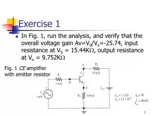
Exercise 1
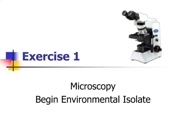
Exercise 1. Microscopy. enables one to study objects too small to be seen and examined with the naked eye an optical instrument consisting of a system of lenses that gives sharp, distinct and highly magnified images of minute objects

Exercise 1
E N D
Presentation Transcript
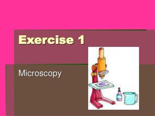
Exercise 1 Microscopy
enables one to study objects too small to be seen and examined with the naked eye • an optical instrument consisting of a system of lenses that gives sharp, distinct and highly magnified images of minute objects • different types depend on usage, source of light, and maximum capacity to enlarge an object • uses ordinary room lighting condition, easier to move about and use
A. Parts and Functions 1. Eyepiece/Ocular – topmost portion • Small, removable tube containing lenses with the magnifying power etched on its surface • Part where the specimen is viewed • Intermediate image projected by the objective is enlarged by the eyepiece. • *Hence, the term compound microscope is derived from the fact that the specimen is magnified twice, first by the objective and second by the eyepiece. The final image formed is a virtual image.
2. Extension/Draw tube – Through this tube, the image is projected over a distance 3. Body tube – wide hollow cylindrical tube which provides a short distance for the image/light to pass through
4. Revolving nosepiece – attached beneath the body tube which serves as the base for one or more objectives. It can be rotated to position the appropriate objective to be used (allows convenient exchange of the objectives)
5. Objectives – small narrow tubes containing compound lenses for magnification a. low power objective (LPO) – shortest, 10x (magnification), 5mm (working distance) b. high power objective (HPO) – 40-43x, 0.46mm c. oil immersion objective (OIO) – requires that oil be placed between the objective lens and the coverslip for a distinct image to form, 100x, 0.13mm d. scanner – allows a wider area of the specimen to be viewed, 2.5x
6. Arm – curved portion connecting the body tube to the base of the microscope; this is where the microscope is held for carrying or tilting; supports the body tube and adjustment knobs; permits adjustment of the stage to a desired angle
7. Adjustment knobs – two pairs of knobs found on both sides of the arm a. Coarse adjustment knob – larger pair; adjust or moves the body tube, together with the objectives, up and down easily. It is used to bring into focus the specimen to be observed. b. Fine adjustment knob – smaller pair; adjusts slowly and is used to sharpen the focus
8. Inclination joint – found at the base of the arm which allows the upper portion of the microscope to be tilted 9. Stage – place where the glass slide (which contains the specimen to be observed) is placed; contains the stage clips and a hole 10. Stage clip – holds the slide in place 11. Opening/Aperture – where light passes through 12. Aperture disc – movable; connected to and beneath the stage contains a series of holes with different sizes for the regulation of the incoming light
13. Substage condenser – used to concentrate the incoming light 14. Iris diaphragm – below the condenser; with a movable lever, also for regulating the incoming light 15. Mirror – found at the base of the microscope which is used to direct the light through the opening of the stage 16. Pillar – region connecting the inclination joint with the stand at the base of the microscope. Together they support and hold the microscope in a steady position 17. Base – keeps the microscope steady at any position of the stage
Parfocal – means that the objectives are optically and mechanically designed so that the distance between the specimen and the aerial image is always constant. Slight refocusing with the aid of fine focus knob is sufficient to restore critical sharpness of the image after changing from objective to another, thus the coarse focus knob need not be operated.
B. Use/Operation of the Microscope • READ the manual!
C. Care of the Microscope • Read the manual…
D. Terms and Concepts in Microscopy • Resolving Power • Limit of resolution • Working distance • Field of Vision • Magnification (Linear) • Parfocal
E. Calibration of the Microscope • *1mm = 1000 micrometers • The ocular micrometer is a glass disc with mounted scale. It is inserted into the eyepiece and must be calibrated for the particular objective, eyepiece and tube length employed before measurements are made. The student microscope has a fixed tube length. • A stage micrometer is a glass slide with graduations of known intervals. The length of one small division is 0.01 mm or 10 micrometers, whereas one big division is 0.1 mm or 100 micrometers.
1. Calibration of the Ocular Micrometer • Use this formula to determine the value of one division on the ocular micrometer: • Calibration factor = SM divisions subtended by OM x Value of one SM div OM division subtended by SM
2. Measurement of the Specimen • The specimen is measured by counting the number of divisions it covers on the ocular micrometer. Knowing the calibration factor, you can compute the size of the specimen in micrometers or millimeters.
F. Techniques for Preparing Specimens for Light Microscopy • Wet mount technique – organism can be observed in its normal living condition. Simple wet mount involves placing on a glass slide a drop of the specimen which is suspended in a fluid. If the specimen is dry, a drop of water is added to the specimen on the glass slide. A coverslip is placed on top of the specimen to prevent drying and to flatten the specimen in order to avoid the refraction of light. • Fixed and Stained preparation
G. Dissecting Microscope • consists of a single lens which provides a large and clear field and gives a magnification of 6x to 20x • for the study of large or thick specimens • useful in examining small organisms and parts of large organisms • gives an erect image
Hypertonic • Hypotonic • Isotonic
Semi-permeability • Phospholipid bilayer
Photosynthesis • Capture solar energy and utilize for the synthesis of organic compounds from water and carbon dioxide in a series of enzyme-mediated complex reaction • Chloroplasts (thylakoid membranes)
Photosynthesis 6 CO2 + 12 H2O Light and chlorophyll---) C6H12O6 + 6 H2O + 6 O2
Four steps 1. Absorption of light (directly light dependent) H2O + 2 NADP+ light---) 2 H+ + 2 NADPH + O2 2. Electron transport (directly light dependent)
3. ATP generation (directly light dependent) 4. Carbon fixation (indirectly light dependent) 6 CO2 + 12 H2O + 18 ATP + 12 NADPH ---) C6H12O6 + 18 ADP + 12 NADP + 6 H+
Respiration • Slow, controlled release of energy through the enzymatic breakdown of organic substances into simpler products • Mitochondria (inner membrane)
Respiration • Aerobic C6H12O6 + 6 H2O + 6 O2 Enzymes-----) 6 CO2 + 12 H2O + Energy
Anaerobic • C6H12O6 Enzymes-----) 2CO2 + C2H5OH (ethanol) + Energy • C6H12O6 Enzymes-----) 2C3H6O3 (lactic acid) + Energy
Cellular Respiration in yeast • Respire both aerobically and anaerobically • Easily handled • If oxygen is available, it will respire both aerobically and anaerobically